Allergies / Pollens


Allergy / Sensitivity


Bacterial Diseases
Bacterial Diseases
Babesia Genus (1-15)
Bacillus Anthracis
This is the etiologic agent of anthrax-a common disease of livestock and, occasionally, of humans-and the only obligate pathogen within the genus Bacillus. B. anthracis is a Gram-positive, endospore-forming, rod-shaped bacterium, with a width of 1.0-1.2 µm and a length of 3-5 µm.
Bordetella Genus (1-39)
These are small (0.2 – 0.7 µm), Gram-negative coccobacilli of the phylum Proteobacteria. Bordetella species, with the exception of B. petrii, are obligate aerobes, as well as highly fastidious, or difficult to culture. All species can infect humans. The first three species to be described (B. pertussis, B. parapertussis, B. bronchiseptica,); are sometimes referred to as the ‘classical species’. One of these (B. bronchiseptica) is also motile.
B. pertussis and occasionally B. parapertussis cause pertussis or whooping cough in humans, and some B. parapertussisstrains can colonise sheep. B. bronchiseptica rarely infects healthy humans, though disease in immunocompromised patients has been reported. B. bronchiseptica causes several diseases in other mammals, including kennel cough and atrophic rhinitis in dogs and pigs, respectively. Other members of the genus cause similar diseases in other mammals, and in birds (B. hinzii, B. avium). Source
Bordetella Pertussis
This is a Gram-negative, aerobic, pathogenic, encapsulated coccobacillus of the genusBordetella, and the causative agent of pertussis or whooping cough…. Its virulence factors includepertussis toxin, filamentous hæmagglutinin, pertactin, fimbria, and tracheal cytotoxin.
Borrelia
The various species of Borrelia are known to humans in the form of Lyme disease and recurring fever, transmitted through tick or flea bite. The cycle of Borrelia through animals is related to the tick’s life cycle. The tick has four stages in its two-year life cycle, egg, larva, nymph and adult. Between each stage the tick needs a blood meal in order to mature. The tick usually acquires the spirochaete during its larval stage, when it feeds on small animals such as rodents or birds. Usually the tick picks up Borrelia from the white-footed mouse, which is commonly infected. The tick then becomes the host for the spirochete. The bacteria resides in the digestive tract of the host for its next nymph and adult stages during which it is passed on to other animals, and sometimes humans.
Lyme disease (named for the town in which it was first identified) can be caused by any number of different species in the genus Borrelia, such as: B. andersonii, B. japonica, B. valaisiana, B. lusitanie, B. turdae. B. tunakii, B. bissettii, and B. lonestari.
Borrelia inhabits the lumen of a tick’s digestive tract. The disease is transmitted to humans from a tick bite when the bacteria migrates up to the ticks salivary glands, and through the opening created by the tick. Ticks increase salivation during gorging, prompting the migration of the saliva from the digestive tract. Because migration from the gut takes a few days, transmission of the disease usually does not happen until after the first 24 hours of attachment.
During early stages of the disease the bacteria is localized in the skin and manifests itself as a characteristic bulls-eye rash, called Erythema Migrans (not in all cases, some people develop no rash). If the disease is caught in this stage and treated, further complications can be avoided. If the disease is not treated, symptoms can include arthritis, cranial neuropathy (specifically facial palsy), and meningitis (abnormal cerebrospinal fluid).
Recurring fever as the result of tick or flea bites have also been traced back to species of the genus Borrelia. More than 20 Borrelia species have been linked with recurring fever, among these is Borrelia recurrentis, which is transmitted by flea bite.
Borrelia (other 1-20)
Borrelia Burgdorferi (lyme)
This is a bacterial species of the spirochete class of the genus Borrelia. B. burgdorferi exists in North America and Europe and is the predominant causative agent of Lyme disease in the United States. Known for the “bullseye” rash.
Brucella
Brucellosis is an infectious disease caused by a type of bacteria called Brucella. The bacteria can spread from animals to humans. There are several different strains of Brucella bacteria. Some types are seen in cows. Others occur in dogs, pigs, sheep, goats, and camels. Recently, scientists have seen new strains in the red fox and certain marine animals, including seals. Brucella in animals cannot be cured.
Brucellosis is rare in the U.S. because of effective animal disease control programs. Fewer than 200 people get sick with the disease each year in the U.S. It is most often seen in the spring and summer months.
Campylobacter Jejuni (C. jejuni)
This infection causes diarrhea, which may be watery or sticky and can contain blood (usually occult) and fecal leukocytes (white cells). Other symptoms often present are fever, abdominal pain, nausea, headache and muscle pain. The illness usually occurs 2-5 days after ingestion of the contaminated food or water.
Chlamydia Psittaci
This is a lethal intracellular bacterial species that may cause endemic avian chlamydiosis, epizootic outbreaks in mammals, and respiratory psittacosis in humans.
Clostridium Botulinum
This is a Gram-positive, rod-shaped, anaerobic, spore-forming, motile bacterium with the ability to produce the neurotoxin botulinum.
Clostridium Difficile (C. difficile)
This is a bacterium that causes diarrhea and more serious intestinal conditions such as colitis.
Clostridium Perfringens (C. perfringens)
This is a spore-forming gram-positive bacterium that is found in many environmental sources as well as in the intestines of humans and animals.
Corynebacterium Diphtheriae
This is a nonmotile, noncapsulated, club-shaped, Gram-positive bacillus. Toxigenic strains are lysogenic for one of a family of corynebacteriophages that carry the structural gene for diphtheria toxin, tox.
E. Coli
This is a bacterium commonly found in the intestines of humans and other animals, where it usually causes no harm. Some strains can cause severe food poisoning, especially in old people and children.
Enterococus Faecalis / Faecium
is a Gram-positive, commensal bacterium inhabiting the gastrointestinal tracts of humans and other mammals. Like other species in the genus Enterococcus, E. faecalis can cause life-threatening infections in humans, especially in the nosocomial (hospital) environment, where the naturally high levels of antibiotic resistance found in E. faecalis contribute to its pathogenicity. E. faecalis has been frequently found in re-infected root canal-treated teeth in prevalence values ranging from 30% to 90% of the cases. Source
Francisella Tularensis (Tularemia)
This is a disease of animals and humans. Rabbits, hares, and rodents are especially susceptible and often die in large numbers during outbreaks. Humans can become infected through several routes, including tick and deer fly bites.
Haemophilus Influenzae
This is a type of bacteria that mainly causes illness in babies and young children. These bacteria can cause infections in people of all ages ranging from mild, such as an ear infection, to severe, such as a bloodstream infection. In spite of the name, H. influenzae do not cause influenza (the “flu”).
Helicobacter Pylori (h. pylori)
This can enter your body and live in your digestive tract. After many years, they can cause sores, called ulcers, in the lining of your stomach or the upper part of your small intestine. For some people, an infection can lead to stomach cancer. Infection with H. pylori is common. About two-thirds of the world’s population has it in their bodies. For most people, it doesn’t cause ulcers or any other symptoms. After H. pylori enters your body, it attacks the lining of your stomach, which usually protects you from the acid your body uses to digest food. Once the bacteria have done enough damage, acid can get through the lining, which leads to ulcers. These may bleed, cause infections, or keep food from moving through your digestive tract. You can get H. pylori from food, water, or utensils. The germs live in the body for years before symptoms start, but most people who have it will never get ulcers.
Legionella Pneumophila (legionellosis)
This is a respiratory disease caused by Legionella bacteria. Sometimes the bacteria cause a serious type of pneumonia (lung infection) called Legionnaires’ disease.
Leptospira Interrogans
This is a Gram negative, obligate aerobe spirochete, with periplasmic flagella. When viewed through a light microscope, it often resembles a question mark, and this gives the species its name. It is a member of the genusLeptospira.
Listeria Monocytogenes
This is the species of pathogenic bacteria that causes the infection listeriosis. It is a facultative anaerobic bacterium, capable of surviving in the presence or absence of oxygen.
Lyme
This is an infectious disease caused by bacteria of the Borrelia type which is spread by ticks. The most common sign of infection is an expanding area of redness on the skin, known as erythema migrans, that begins at the site of a tick bite about a week after it has occurred. The rash is typically neither itchy nor painful. Approximately 25–50% of infected people do not develop a rash. Other early symptoms may include fever, headache and feeling tired.If untreated, symptoms may include loss of the ability to move one or both sides of the face, joint pains, severe headaches with neck stiffness, or heart palpitations, among others. Months to years later, repeated episodes of joint pain and swelling may occur. Occasionally, people develop shooting pains or tingling in their arms and legs. Despite appropriate treatment, about 10 to 20% of people develop joint pains, memory problems, and feel tired for at least six months. Source
MRSA
This is methicillin-resistant Staphylococcus aureus, a type of staph bacteria that is resistant to several antibiotics. In the general community, MRSA most often causes skin infections. In some cases, it causes pneumonia (lung infection) and other issues. If left untreated, MRSA infections can become severe and cause sepsis–a life-threatening reaction to severe infection in the body. In a healthcare setting, such as a hospital or nursing home, MRSA can cause severe problems such as bloodstream infections, pneumonia and surgical site infections.
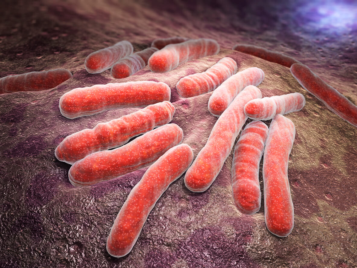
Mycobacterium Tuberculosis
This is an obligate pathogenic bacterial species in the family Mycobacteriaceae and the causative agent of tuberculosis.
Mycoplasma Genus (1-5)
This is a genus of bacteria that lack a cell wall around their cell membrane. Without a cell wall, they are unaffected by many common antibiotics such as penicillin or other beta-lactam antibiotics that target cell wall synthesis. They can be parasitic or saprotrophic. Several species are pathogenic in humans, including M. pneumoniae, which is an important cause of atypical pneumonia and other respiratory disorders, and M. genitalium, which is believed to be involved in pelvic inflammatory diseases. Mycoplasma species are the smallest bacterial cells yet discovered.
Neisseria Meningitidis
This is often referred to as meningococcus, is a Gram-negative bacterium that can cause meningitis and other forms of meningococcal disease such as meningococcemia, a life-threatening sepsis.
Pseudomonas Aeruginosa
Serious infections from P. aeruginosa generally occur only in healthcare (nosocomial) settings, but people can also develop mild infections in other environments.
Rickettsia Rickettsii
This is the small, aerobic gram-negative bacterium that is the cause Rocky Mountain spotted fever in humans (and other vertebrates).
Salmonella infection (salmonellosis)
This is a bacterial disease of the intestinal tract Salmonella is a group of bacteria that causes typhoid fever, food poisoning, gastroenteritis, enteric fever and other illnesses. People become infected mostly through contaminated water or foods, especially meat, poultry and eggs.
Shigella Sonnei
This is a non-motile, nonspore-forming, facultative anaerobic Gram-negative bacterium. Its non-motile characteristic means that this species doesn’t have flagella to facilitate its movement like many other human enterobacteria.
Staphylococcus Aureus
Staphylococcus (sometimes called “staph”) is a group of bacteria that can cause a multitude of diseases. Staph infections may cause disease due to direct infection or due to the production of toxins by the bacteria.
Streptococcus Group A (group A strep)
This is a type of bacterium that can cause many different infections that range from minor illnesses to very serious and deadly diseases.
Streptococcus Group B (group B strep)
This is a common bacterium often carried in your intestines or lower genital tract. Group B strep is usually harmless in adults. In newborns, however, it can cause a serious illness known as group B strep disease.
Ureaplasma Genus (1-2)
Ureaplasma biovars, Ureaplasma urealyticum and Ureaplasma parvum, are now designated as separate species. Separation of these species is not possible except via molecular techniques such as polymerase chain reaction (PCR). Therefore, they are now considered together as Ureaplasma species. U parvum is generally the most common species detected in various clinical specimens but U urealyticum is apparently more pathogenic in conditions such as male urethritis. This differential pathogenicity at the species level has not been shown consistently for other disease conditions.
Fungus
Fungus
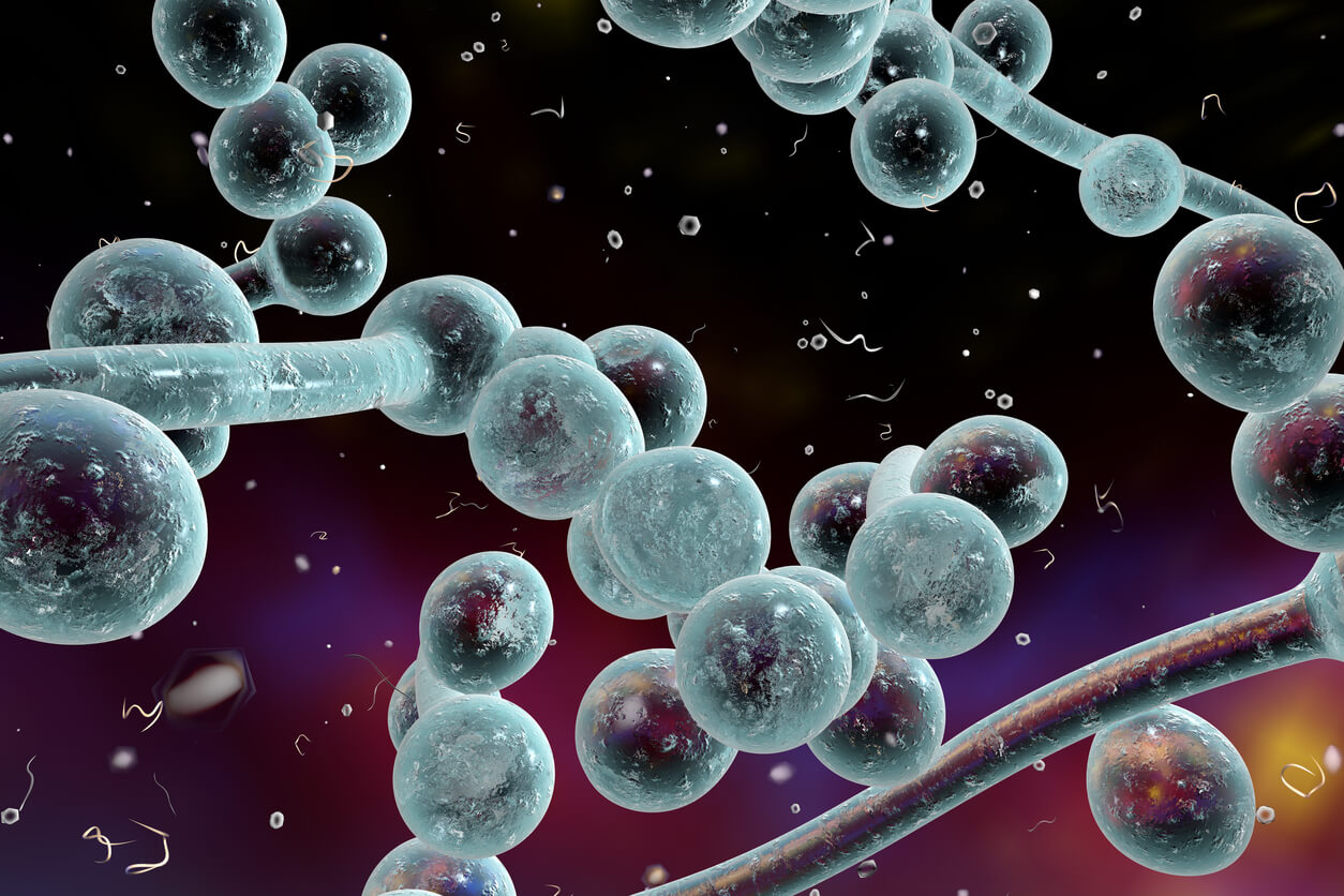
There are over 40 different species of Candida Yeast, however, only a handful are harmful to us. These are the types that interact with humans. Chronic out of balance candida and other forms of gut dysbiosis can lead to “leaky gut” syndrome which is very simply, the inflammation and weakening of the intestinal walls.
The Candida cells, which are relatively benign in their yeast form, adopt their fungal form and begin to grow hyphae – the long branches that grow out of the fungus. These branches invade the cells in your intestinal lining, creating inflammation and permeating the membrane that prevents substances from leaking out.
This condition is characterized by leaky wall of the guts that makes it possible for different substances to penetrate into the body such as microbes, undigested foods, toxins and many others. They are supposed to stay inside the gut, but when they penetrate the body, they can cause dangerous health disorders.
The most common disorders caused by leaky gut are: obesity, schizophrenia, type 1 and 2 diabetes, irritable bowel syndrome, celiac disease, Chrohn’s disease and rheumatoid arthritis. The digestion is the first affected by leaky gut which is manifested as inflammatory gut disease like ulcerous colitis or irritable bowel syndrome.
Moreover, it can be manifested as allergy or sensitivity to some ingredients and as a result of that, it can cause pain and other digestive problems.
Leaky gut can also result in stiffness, pain and difficulty moving. You can experience pain in other parts of your body as well due to leaky gut. This disorder can also cause psoriasis and eczema, but it can be manifested as rosacea acne as well. Leaky gut can weaken your immune system which will lead to constant colds and sinus infections. It affects your brain as well, and people who have experienced this disorder, report a feeling of tiredness, anxiety and depression.
If you experience some of the above mentioned symptoms, make sure to consult your doctor and do the necessary tests to discover whether you are dealing with leaky gut syndrome or not.
Make sure to avoid the consumption of sugar, lactose and gluten since they are considered to be the heaviest food that trigger leaky gut disorder. Instead, you should consume coconut products, fermented foods such as pickles and veggies, bone marrow soup and raw dairy products.
Nowadays, people are under a lot of stress which is the main factor for all kinds of health issues because it weakens your immune system which can lead to leaky gut.
In order to relieve stress, try to exercise more, spend some quality time with people you love, meditate or write a personal dairy.
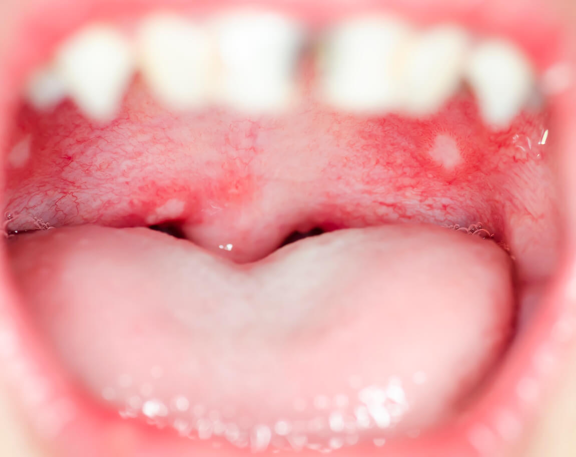
Candida Albicans
This species is the most common species of yeast in our bodies. It lives in our digestive tract and on our skin. Normally it lives in our bodies in a commensal relationship with us meaning we both benefit from each other. However, candida ablicans overgrowth can occur which results in infection most commonly in the genital area and the mouth.
Candida Glabrata
Candida Krusei
Candida Lusitaniae
This species was first identified to cause a yeast infection in 1979 but very few cases were reported until recently with wide use of procedures such as chemotherapy and bone marrow transplants which leaves humans open to infection by this species.
Candida Parapsilosis
Candida Stellatoidea
Candida Tropicalis
A species of mitosporic fungi that is a major cause of septicemia and disseminated candidiasis especially in patients with lymphoma, leukemia and diabetes mellitis. It is also found as part of the normal human mucocutaneous flora.
Cryptococcus Gattii
Cryptococcus gattii causes the human diseases of pulmonary cryptococcosis (lung infection), basal meningitis, and cerebral cryptococcomas. Occasionally, the fungus is associated with skin, soft tissue, lymph node, bone, and joint infections. Source
Mucormycosis
This is any fungal infection caused by fungi in the order Mucorales. Generally, species in the Mucor,Rhizopus, Absidia, and Cunninghamella genera are most often implicated.
The disease is often characterized by hyphae growing in and around blood vessels and can be potentially life-threatening in diabetic or severely immunocompromised individuals. Source
Pneumocystis Jiroveci
This is a yeast-like fungus of the genus Pneumocystis. Source
Sporotrichosis
Widespread infectious disease marked by nodules or ulcers of theskin, chiefly affecting humans and domestic mammals and caused bythe fungus Sporothrix schenckii. Source
Talaromyces
This is a genus of fungi in the family Trichocomaceae. Described in 1955 by American mycologist Chester Ray Benjamin, species in the genus form soft, cottony fruit bodies (ascocarps) with cell walls made of tightly interwoven hyphae. The fruit bodies are often yellowish or are surrounded by yellowish granules. A 2008 estimate placed 42 species in the genus, but several new species have since been described. Source
Tinea Corporis
This is a superficial fungal infection (dermatophytosis) of the arms and legs, especially on glabrous skin; however, it may occur on any part of the body.
Signs and Symptoms
It may have a variety of appearances; most easily identifiable are the enlarging raised red rings with a central area of clearing (ringworm). The same appearances of ringworm may also occur on the scalp (tinea capitis), beard area (tinea barbae) or the groin (tinea cruris, known as jock itch or dhobi itch).
Other classic features of tinea corporis include:
The edge of the rash appears elevated and is scaly to touch.
Sometimes the skin surrounding the rash may be dry and flaky.
Almost invariably, there will be hair loss in areas of the infection. Source
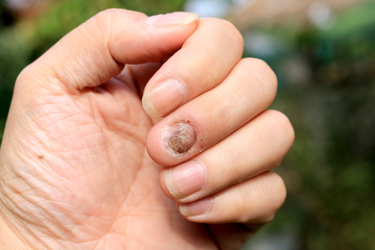
Tinea Unguium (Onychomycosis)
This is a fungal infection of the nail. This condition may affect toenails or fingernails, but toenail infections are particularly common. Source
Human Toxins
Alcohol
Excessive alcohol use can cause the pancreas to produce toxic substances that interfere with proper functioning. The resulting inflammation is called pancreatitis, a serious problem that can destroy the pancreas. One of the most frequent causes of chronic pancreatitis is alcohol abuse. The liver’s job is to break down harmful substances, including alcohol. Excessive drinking can cause alcoholic hepatitis which can lead to the development of jaundice (yellowing of the skin and eyes).
Chronic liver inflammation can lead to severe scarring known as cirrhosis. This formation of scar tissue can destroy the liver. When the liver fails to perform, toxic substances remain in your body. Alcoholic liver disease is the liver manifestations of alcohol overconsumption, including fatty liver (buildup of fat in the liver. Its normal to have fat in your liver, however more than 5 – 10 % is not normal and you may have fatty liver), alcoholic hepatitis (an inflammatory condition of the liver because of drinking too much alcohol for a very long time), and chronic hepatitis (inflammation of the liver) with liver fibrosis (the first stage of liver scarring,)or cirrhosis(which is scar tissue that replaces the normal tissue of the liver).
When the pancreas and liver don’t function properly, the risk of hypoglycemia (low blood sugar) rises. A damaged pancreas can cause the body to be unable to utilize sugar due to a lack of insulin, which can lead to hyperglycemia. Unbalanced blood sugar levels can be a dangerous problem, especially for people with diabetes.
Drinking also releases excess GABA and dopamine, two naturally occurring neurotransmitters. GABA is responsible for calming the brain down, and dopamine is responsible for pleasure, a part of the brain’s reward system. Too much of these neurotransmitters can lead to shortness of breath, high blood pressure, increased heart rate, night terrors, delusions, hallucinations, spasms, and increased levels of both aggression and depression.
Chemical Toxicity
According to the United States Environmental Protection Agency or EPA, the most important threat to both human, animal and plant life on earth comes from the effects of toxic chemicals. Hundreds of thousands of chemicals have been produced in the world in the past two hundred years, especially, often with little understanding of their toxicity – until a problem arises. Thousands of toxic chemicals have found their way into our air, food and water supplies worldwide. No place on earth is free of them anymore because they are carried by the wind and the rain to every corner of the earth. Studies reveal that everyone in the developed nations has hundreds of these toxic chemicals in their blood and stored in their body tissues. Toxic chemicals contribute to every possible type of physical and mental health problem imaginable.
One important group of chemicals, the endocrine disruptors. These are toxic chemicals that disrupt the hormone systems of plants, animals and human beings. Most people have at least a dozen of these inside the body, and many people today are born with them, having acquired them from your mother in utero.
If you get enough of these chemicals in your body, they can wreak havoc on your hormones due to the following mechanisms:
– Increasing production of certain hormones
– Decreasing production of other hormones
– Imitating hormones
– Turning one hormone into another
– Interfering with hormone signaling
– Telling cells to die prematurely
– Competing with essential nutrients
– Binding to essential hormones
– Accumulating in organs that produce hormones.
Some of the worst are:
BPA. This chemical mimics estrogen in the body.
Dioxin. This is a pesticide sprayed on some food products.
Atrazine. This is a toxic herbicide sprayed on some food products, especially corn.
Phthalates. This is another toxic chemical that can signal cells to die. It is found in some plastic food containers, some personal care products, and food wrap that says recycle #3.
Perchlorate. This chemical is sometimes used to kill germs that live in tap water. It interferes with iodine in the body.
Other iodine antagonists. Other chemicals that interfere with iodine in the body and thereby cause thyroid problems are bromine and fluorine.
Fire retardants (PBDEs). These are very persistent chemicals that imitate thyroid hormones and contribute to thyroid problems.
Perfluorinated chemicals. These are chemicals used to make non-stick coatings on pots and water-resistant coatings for clothing. They are very persistent chemicals that have many negative effects on the body including kidney and thyroid disease, low birth weight, damaged sperm and high cholesterol. Most people have some in their bodies because they do not degrade.
Organophosphates. These are very toxic pesticides used to stop insect reproduction.
Glycol ethers. These are endocrine-disrupting chemicals found in solvents used in paints, brake fluid, cleaning products and cosmetics. They can cause asthma, allergies and blood abnormalities, among other problems.
Arsenic, lead, copper and mercury. These toxic metals are also considered endocrine disrupters. Copper and mercury affect the thyroid gland. Lead and arsenic – widely used in pesticides – can affect many glands in the body.
Medical drugs in the water. Unfortunately, some medical and over-the-counter drugs do not break down or biodegrade quickly.
Drugs, Medication
A person with drug toxicity has accumulated too much of a medication in the bloodstream. The effects of the medication are more pronounced at toxic levels, and side effects may be severe. Toxicity may result when the dose is too high, or it may result when the liver or kidneys are unable to remove the drug from the bloodstream. Many commonly prescribed medications can accumulate in the bloodstream and result in toxicity.
Symptoms of drug toxicity depends on the drug taken. Symptoms of drug toxicity can be broken down into: Symptoms of GHB abuse which include: palpitations, confusion, lethargy, coma, low blood pressure, low body temperature, muscle spasms, slow breathing, slow heart rate, vomiting, violent behavior. Others include symptoms of hallucinogens, symptoms of narcotics, symptoms or sedatives, and symptoms of stimulants.
Drugs, Recreational
Recreational drugs are chemical substances that are taken for enjoyment instead of medical reasons. Psychedelic Mushrooms, Amphetamines, Ecstasy, Cocaine, Opium, Heroin and Cannabis are just some of recreational drugs out there. They can lead to addiction, health and social problems and crime. Most recreational drugs are illegal.
Electromagnetic Radiation
This is a kind of radiation including visible light, radio waves, gamma rays, and X-rays, in which electric and magnetic fields vary simultaneously. Sunlight is also a form of EM energy, but visible light is only a small portion of the EM spectrum, which contains a broad range of electromagnetic wavelengths. The study of electromagnetism deals with how electrically charged particles interact with each other and with magnetic fields.
There are four main electromagnetic interactions:
The force of attraction or repulsion between electric charges is inversely proportional to the square of the distance between them.
Magnetic poles come in pairs that attract and repel each other, much as electric charges do.
An electric current in a wire produces a magnetic field whose direction depends on the direction of the current.
A moving electric field produces a magnetic field, and vice versa.
EM radiation is created when an atomic particle, such as an electron, is accelerated by an electric field, causing it to move. The movement produces oscillating electric and magnetic fields, which travel at right angles to each other in a bundle of light energy called a photon. Photons travel in harmonic waves at the fastest speed possible in the universe: 186,282 miles per second (299,792,458 meters per second) in a vacuum, also known as the speed of light. The waves have certain characteristics, given as frequency, wavelength or energy.
Excitotoxins
Excitotoxins are a class of chemicals (usually amino acids) that overstimulate neuron receptors. Neuron receptors allow brain cells to communicate with each other, but when they’re exposed to excitotoxins, they fire impulses at such a rapid rate that they become exhausted. Several hours later, these depleted neurons die. Excitotoxins can cross the placental barrier, possibly harming the brains of unborn children. Excitotoxins also cross the blood brain barrier and are known to cause migraines, seizures, neurological disorders, blurred vision, increased appetite, overeating, infertility and reproductive disorders, impaired brain function, cancer, and heart and cardiovascular damage.
Excitotoxicity is the pathological process by which nerve cells are damaged or killed by excessive stimulation by neurotransmitters such as glutamate and similar substances. This occurs when receptors for the excitatory neurotransmitter glutamate (glutamate receptors) such as the NMDA receptor and AMPA receptor are overactivated by glutamatergic storm. Excitotoxins like NMDA and kainic acid which bind to these receptors, as well as pathologically high levels of glutamate, can cause excitotoxicity by allowing high levels of calcium ions (Ca2+) to enter the cell. calcium ionsenter into cells activates several enzymes, including phospholipases, endonucleases, and proteases such as calpain. These enzymes go on to damage cell structures such as components of the cytoskeleton, membrane, and DNA. Examples of excitotoxins include: aspartame (NutraSweet), sucralose, cysteine, hydrolyzed protein, aspartic acid and food coloring. MSG is one of the worst and is disguised under at least 30 other names which include: autolyzed yeast, calcium caseinate, gelatin, glutamate, glutamic acid, hydrolyzed protein, monopotassium glutamate, monosodium glutamate, sodium caseinate, textured protein, yeast extract, yeast food, and yeast nutrient.
Hair Dyes
In certain individuals, the use of hair coloring can result in allergic reactions and/or skin irritation. Individuals allergic to gluten for example, will need to be cautious when purchasing hair color since certain hair dye includes gluten. Gluten does not need to be ingested for it to cause an allergy. Skin contact with gluten may cause a reaction; therefore, leading to an allergy. Symptoms of these reactions can include redness, sores, itching, burning sensation and discomfort. Symptoms will sometimes not be apparent immediately following the application and processing of the tint, but can also arise after hours or even a day later. Source
Herbicide Toxicity
Although many modern herbicides are less toxic than their predecessors, they are still poisons and should always be handled with caution.Skin irritations are some of the most common effects when a person comes into contact with herbicides, and are most likely to happen on exposed areas, such as the hands and forearms. Some chemicals may burn the skin and should be washed off immediately with cold water.
Glyphosate, the active ingredient in Monsanto’s Roundup herbicide, is possibly “the most important factor in the development of multiple chronic diseases and conditions that have become prevalent in Westernized societies.” Glyphosate residues are found in most commonly consumed foods in the Western diet courtesy of GM sugar, corn, soy, and wheat.
Research suggests that glyphosate may “enhance the damaging effects of other food-borne chemical residues and toxins in the environment to disrupt normal body functions [including gut bacteria] and induce disease.” Glyphosate causes extreme disruption of the microbe’s function and lifecycle. What’s worse, glyphosate preferentially affects beneficial bacteria, allowing pathogens to overgrow and take over, including the highly toxic Clostridium botulinum.
Glyphosate may stimulate hormone-dependent cancers even at extremely low “environmentally relevant” amounts.
Metabolic Waste
Metabolic toxins (or body toxins) are normal by-products of your metabolism occurring throughout the mind and body. Organic chemist Ludwig Brieger defined a toxin as a poisonous substance produced within living cells or organisms. This excluded manufactured substances (chemical) created by artificial processes. Simply put, metabolic toxins are toxic waste byproducts produced throughout every metabolic pathway in the mind and body that must be eliminated from the body. Numerous factors may contribute toward an excess of metabolic toxins. However, the two most common factors today include:
1. Nutrient imbalances
Nutrient imbalances include nutrient excesses and deficiencies, inherited enzyme deficits, toxic elements, chemical toxicants, medications, stress, and so on. Metabolic toxins can produce a long list of symptoms and conditions throughout the mind and body.
Each metabolic pathway (urea cycle, citric acid cycle, carbohydrate metabolism, neurotransmitter metabolism, etc.) requires a specific combination and proper amount of essential nutrients (or derivatives) during each step of the process.
If there is a deficiency or excess of any of the nutrients required for the specific metabolic pathway, the pathway is not completed in an efficient manner and results in an excess of a metabolic toxin. In any pathway, combinations of essential nutrients may be synthesized to produce additional metabolites required for the metabolic pathway to function properly.
Metabolic toxins produce a variety of metabolic intermediates known as organic acids. A urinary organic acids lab analysis is a functional analysis. This simply means the organic acids analysis can be helpful for determining whether a sufficient amount of a particular nutrient is available for a variety of metabolic pathways. If a particular nutrient is deficient, it produces specific metabolic intermediates or metabolic toxins.
2. Sugar and simple carbohydrates
Sugar, in its variety of incognito names such as high fructose corn syrup, agave, aspartame, and so on, are the most common contributors for metabolic toxins. Sugar also contributes toward nutrient imbalances further increasing metabolic toxins. Simply put, sugar is poison to the mind and body.
Simple carbohydrates such as refined flour (white bread, pasta, white rice, etc.) are all major contributors.
Paint
This is any liquid, liquefiable, or mastic composition that, after application to a substrate in a thin layer, converts to a solid film. It is most commonly used to protect, color, or provide texture to objects. Paint can be made or purchased in many colors—and in many different types, such as watercolor, synthetic, etc. Paint is typically stored, sold, and applied as a liquid, but most types dry into a solid. Source
Pesticide Toxicity
Pesticide poisoning symptoms are similar to those of other illnesses and poisonings. Unfortunately, all pesticide poisoning symptoms are not the same. Each chemical family (i.e., organophosphates, carbamates, chlorinated hydrocarbons) can attack the human body in a different way. However, you should be aware of the general symptoms of pesticide poisoning.
Mild Poisoning or Early Symptoms of Acute Poisoning include: headache, fatigue, weakness, dizziness, restlessness, nervousness, perspiration, nausea, diarrhea, loss of appetite, loss of weight, thirst, moodiness, soreness in joints, skin irritation, eye irritation, irritation of the nose and throat.
Moderate Poisoning or Early Symptoms of Acute Poisoning: nausea, diarrhea, excessive saliva, stomach cramps, excessive perspiration, trembling, no muscle coordination, muscle twitches, extreme weakness, mental confusion, blurred vision, difficulty in breathing, cough, rapid pulse, flushed or yellow skin, weeping.
Severe or Acute Poisoning: fever, intense thirst, increased rate of breathing, vomiting, uncontrollable muscle twitches, pinpoint pupils, convulsions, inability to breathe, unconsciousness.
Smog Pollution
Exposure to smog can lead to several different types of short-term health problems due to its ozone content. These include:
Coughing and throat or chest irritation: High levels of ozone can irritate your respiratory system, generally lasting for a few hours after you’ve been exposed to smog. However, ozone can continue to harm your lungs even after symptoms disappear.
Worsening of asthma symptoms: If you suffer from asthma, exposure to high levels of ozone from smog can trigger asthma attacks.
Difficulty breathing and lung damage: Smog can make it feel difficult to breathe deeply, especially during exercise, according to the Mayo Clinic. This is because of the effects of ozone on lung function.
It’s important to note that smog affects everyone differently, and some people are more susceptible to its negative effects. Children, seniors, and people with asthma need to be especially careful on smoggy days.
Smoke
This is a collection of airborne solid and liquid particulates and gases emitted when a material undergoes combustion or pyrolysis, together with the quantity of air that is entrained or otherwise mixed into the mass. It is commonly an unwanted by-product of fires (including stoves, candles, oil lamps, and fireplaces), but may also be used for pest control (fumigation), communication (smoke signals), defensive and offensive capabilities in the military (smoke screen), cooking, or smoking(tobacco, cannabis, etc.). Smoke is used in rituals where incense, sage, or resin is burned to produce a smell for spiritual purposes. Smoke is sometimes used as a flavoring agent, and preservative for various foodstuffs. Smoke is also a component of internal combustion engine exhaust gas, particularly diesel exhaust. Source
Stimulants
This is an overarching term that covers many drugs including those that increase activity of the body, drugs that are pleasurable and invigorating, or drugs that have sympathomimetic effects. The term stimulant encompasses a broad category of substances, including those prescribed for medical conditions; those manufactured for illicit substance abuse; and those found in over-the-counter (OTC) decongestants, herbal extracts, caffeinated beverages, and cigarettes. The symptoms of a sublethal stimulant overdose may include dizziness, tremor, irritability, confusion, hostility, hallucinations, panic, headache, skin flushing, chest pain, palpitations, cardiac arrhythmias, hypertension, vomiting, cramps, and excessive sweating.
Stress Hormones
Tobacco/Nicotine
Vaccinations
Information is from these sites:
http://www.healthline.com/health/alcohol/effects-on-body
http://www.quitalcohol.com/the-truth-about-what-alcohol-does-to-your-body.html
https://www.fda.gov/drugs/resourcesforyou/consumers/ucm143566.htm
http://www.livescience.com/38169-electromagnetism.html
http://www.prevention.com/mind-body/how-lower-cortisol-manage-stress
https://quitday.org/smoking-effects/nicotine-poisoning/
http://healthliteracy.worlded.org/docs/tobacco/Unit1/1what_is.html

Minerals: Heavy Metals
Aluminum
Foods such as baking powder, self rising flour, salt, baby formula, coffee creamers, baked goods and processed foods, coloring and caking agents.
Vaccines-Hepatitis A and B, Hib, DTaP (diphtheria, tetanus, pertussis), pneumococcal vaccine, Gardasil (HPV), and others.
Removing mercury from vaccines and replacing it with aluminum may be increasing the problems from BOTH toxins in your body. The reason for this is because aluminum impairs your body’s ability to excrete mercury by impeding your glutathione production. Glutathione is your most important intracellular detoxifier, required for reversing oxidative stress. So, if your aluminum load is high, your body will potentially become more toxic from the mercury from, say, flu shots and fish because you are now on “aluminum overload” and your detoxification system no longer functions well.
The best way to protect yourself is to be careful about your choices in food and personal products, and minimize your use of vaccines and other drugs that are often contaminated with aluminum.
Antimony
Antimony toxicity occurs either due to occupational exposure or during therapy. Occupational exposure may cause respiratory irritation, pneumoconiosis, antimony spots on the skin and gastrointestinal symptoms. In addition, antimony trioxide is possibly carcinogenic to humans. Because antimony is found naturally in the environment, the general population is exposed to low levels of it every day, primarily in food, drinking water, and air. Exposure to antimony at high levels can result in a variety of adverse health effects. Breathing high levels for a long time can irritate the eyes and lungs and can cause heart and lung problems, stomach pain and ulcers, diarrhea, and vomiting.
Arsenic
Arsenic is a heavy metal which is a natural component of the earth’s crust. It exists in compounds that may be organic or inorganic. It is highly toxic in its inorganic form. Poisoning can occur by ingestion, inhalation and dermal absorption. Elemental arsenic is the least toxic. Trivalent arsenic is well absorbed through the skin and is 60 times more toxic than pentavalent arsenic, which is well absorbed by the gut. Arsine gas is highly toxic. Regular exposure leads to cancer and other toxic health effects, including cardiovascular disease, skin hyperpigmentation, keratoses, neurological problems, and developmental disorders. Toxicity is due to arsenic’s effect on many cell enzymes, which affect metabolism and DNA repair. Arsenic poisoning symptoms begin with nausea, vomiting, abdominal pain, and severe diarrhea. Arsenic is excreted in urine but can also accumulate in many body tissues.
Barium
Barium carbonate is relatively insoluble in water, it is toxic to humans because it is soluble in the gastrointestinal tract. Barium is a soft, silvery-white metal. It is an active metal, reacting with air, water, acids and bases. Because it is insoluble in the body, barium sulfate is used as an x-ray tracer for the stomach and intestines. Barium is also used in drilling fluids for oil exploration, as well as in paints, fireworks (where it produces a green color), glass and rubber making. It is also used in water softeners, desiccants and rodent poisons. Barium It is never found in nature as a free element. Barium exposure can happen through a number of channels including occupational exposure, groundwater contamination, environmental pollution, cigarette smoke, and certain medical procedures as mentioned above. Industrial use of Barium is perhaps of the largest concern due to the potential for massive environmental pollution.
Bismuth
While many people will tell you that bismuth is non-toxic in small amounts, sufficient exposure can produce nausea, headache, diarrhea, and pain. According to the Department of Physiology at the University of Tübingen in Germany, anemia is another potential negative side effect of exposure to bismuth and caution is advised when taking any medication containing bismuth. Certain metals are known to reduce sperm metabolism and contribute to infertility in men. Bismuth has been suspected to be one of those metals.
It is a naturally occurring metal used to manufacture solder, fishing anchors, shotgun pellets, and more. It is found naturally in very small amounts in some foods and its sulfide and oxide compounds are important for use in cosmetics and medicines. Bismuth is not available as a supplement because it is not essential to your body. Bismuth doesn’t provide any nutritional benefits directly, although it can be of help with gastrointestinal disorders, which is why it is used in brand-name products such as Pepto-Bismol and Kaopectate but consuming too much bismuth can lead to side effects, so consult with your doctor before using it.
Cadmium
This is an extremely toxic metal commonly found in industrial workplaces. It is of no use to the human body and is toxic even at low levels. The negative effects of cadmium on the body are numerous and can impact nearly all systems in the body, including cardiovascular, reproductive, the kidneys, eyes, and even the brain. Exposure can occur if you smoke cigarettes or breathe second- or third-hand cigarette smoke. You can be exposed if you eat foods that contain high levels of cadmium, such as shellfish, liver, and kidney meats. Other foods that contain cadmium are grain cereal products, potatoes, and some leafy vegetables. Cadmium has a very detrimental effect on the central nervous system, including decreased attention and memory in humans. This is likely because cadmium induces neuron cell death. Neurons are brain cells that communicate and transmit information, if they are affected, so is brain function. Cadmium is well-known to cross the placenta and to accumulate in fetal tissues. Prenatal exposure is a threat to the developing brain and results in reduced birth weight and birth size.
Chromium
Chromium hexavalent is a carcinogen that attacks your lungs when inhaled and has been connected to sinus, nasal, and lung cancer. Exposure has been linked to immunity disorders, neuropsychiatric disorders, atherosclerosis, neurodegenerative disorders, congenital disorders, DNA damage, and disruption of bodily processes. In Russia, exposure to chromium hexavalent is widely blamed for premature senility.
Chromate dusts and acids can permanently damage your eyes if they come into direct contact, and other kinds of skin contact may lead to allergic dermatitis, corrosion, skin irritation, sensitization, and even ulcers.
Chromium hexavalent is extremely reactive with vitamin C. When exposure is coupled with vitamin C in the body, it can result in severe damage to DNA inside the lung’s cells. However, outside of the cells, vitamin C actually serves to protect against the damage to the cells. Small amounts of chromium in one form is actually good for people. It makes insulin work better and helps our metabolism.
Cobalt
Cobalt can accumulate to toxic levels in the liver, kidney, pancreas, and heart, as well as the skeleton and skeletal muscle. Cobalt has been found to produce tumors in animals and is likely a human carcinogen as well. Cobalt is naturally occurring element that does have beneficial applications. For instance, cobalt is an essential component of vitamin B12. Cobalt has been added to pigments to produce a distinct blue color. Lithium ion batteries contain cobalt. In the medical field, cobalt-60 is used in radiotherapy and for sterilizing medical equipment. Hip replacements are also made of cobalt. A deficiency of cobalt, which is very rare, can lead to pernicious anemia.
Industrial plants may leak cobalt and other toxic metals into the environment. Once cobalt particles enter the atmosphere, they settle to the ground and enter the food and water supply; most of the population is exposed to cobalt through food, water, and air. Cobalt makes its way through the environment and cannot be destroyed.
Copper
Copper Toxicity is a condition that is increasingly common in this day and age, due to the widespread occurrence of copper in our food, copper fungicides, e-cigs, Copper IUD’s, hot water pipes, along with the common nutritional deficiencies in Zinc, Manganese and other trace minerals that help keep levels of Copper in balance.
Birth control pills increases a woman’s risk of having a Copper toxicity condition due to the effect that estrogen has on the body, increasing copper retention in the kidneys. Estrogen stimulates similar receptors to Aldosterone receptors in the kidneys, increasing Sodium, Copper and water retention. Both estrogens and Aldosterone can increase swelling, Cyst formation, increasing the blood volume which can cause hypertension, stroke, or death if the Liver and Adrenal glands are not able to regulate these hormones in the body.
Copper builds up in the soft tissues of Liver and disrupts the Liver’s metabolic abilities to detoxify and cleanse the blood in general. Copper toxicity in the liver is therefore disrupting to the Liver’s Glucoronidation pathway, that helps to eliminate excessive amounts of Estrogen by making it water soluble. Other toxic heavy metals like Lead, Mercury, Aluminum, and Cadmium will also buildup in the Soft tissues, as a result of Copper competing with Zinc in many enzymes and binding sites in the body.
When Zinc gets displaced by Copper, there will be a reduction in Metallotionein production, which is the main heavy metal binding protein in the body. The production of the body’s main detoxifying agent and antioxidant, Glutathione will also decline when too much Copper gets stored in the Liver organ’s tissues.
Other sources of chemicals which mimic estrogen, known as xeno-estrogens, may also increase the retention of copper. These include pesticides, plastic bags, Volatile organic compounds (VOC’s), growth hormones used on animals, and all petrochemical waste products used in the manufacturing of plastic, gasoline and other petrochemical derivatives. These are all referred to as Xeno-estrogens.
Copper is a very stimulating mineral to the nerves and nervous system. Copper increases the production of epinephrine, norepinephrine, and dopamine while also implicated in a decrease of histamine. These effects on neurotransmitter levels can give rise to many psychological imbalances such as mood swings, depression, mental agitation, feeling over-stimulated, restlessness, anxiety, insomnia and a racing mind with too many thoughts are all hallmarks of elevated Copper toxicity.
Elevated Copper in the body acts like caffeine or even amphetamines. It constantly keeps the conversion of dopamine into norepinephrine going so that you have a constant adrenaline rush to help you be on the go, but you also are unable to settle down or turn off your mind.
Copper toxicity symptoms
•Acne
•Allergies
•Hair loss
•Anemia
•Anorexia
•Anxiety
•Attention deficit disorder
•Arthritis
•Asthma
•Autism
•Candida overgrowth
•Depression
•Dysmenorrhea
•Male infertility
•Prostatitis
•Fibromyalgia
•Migraine Headaches
•PMS
•Chronic infections
•Insomnia
•Racing thoughts
•Neuralgia (nerve pain)
•Sciatica
•Hypertension
•Hypothyroidism
•Schizophrenia
•Bipolar (Manic Depression)
Copper is a necessary component in the manufacturing of ATP (Adenosine triphosphate) which is cellular energy. Low levels of Copper is associated with chronic fatigue. When someone has a Copper toxicity condition, they will most likely also have a concurrent Copper deficiency due to a bio-unavailability. Source
Gold
Gold Toxicity is the toxic effect of gold that occurs when gold is administered to the body. It is usually given for rheumatoid arthritis (RA), juvenile rheumatoid arthritis (JRA), or psoriatic arthritis.
Gold is generally administered to reduce joint pain and joint swelling. In many, gold treatment helps in decreasing joint deformity and joint disability. Although, in about 50% of the individuals, the injections may not be an effective treatment tool. Individuals with the genotype HLA-DR3 have a higher risk for gold therapy-induced Gold Toxicity. In such individuals, kidney toxicity and platelet dysfunction may occur. Gold Toxicity long-term effects may include liver inflammation, blue-grey skin color, and mouth ulcers. There can also be bone marrow suppression resulting in frequent infections. Stopping or discontinuing the use of gold therapy is the first line of treatment for Gold Toxicity. The treatment for arthritis using gold may be resumed, if the side effects improve and go away. The prognosis of Gold Toxicity is generally good with appropriate early diagnosis and treatment including stoppage of the causative gold therapy.
Iron
The body normally absorbs less iron if its stores are full, but some individuals are poorly defended against iron toxicity. Once considered rare, iron overload has emerged as an important disorder of iron metabolism.
Iron overload is known as hemochromatosis and usually is caused by a gene that enhances iron absorption. Other causes of iron overload include repeated blood transfusions, massive doses of dietary iron and rare metabolic disorders. Additionally, long-term overconsumption of iron may cause hemosiderosis, a condition characterized by large deposits of the iron storage protein hemosiderin in the liver and other tissues.
Iron overload is most often diagnosed when tissue damage occurs, especially in iron-storing organs such as the liver. Infections are likely to develop because bacteria thrive on iron-rich blood. Ironically, some of the signs of iron overload are analogous to those of iron deficiency: fatigue, headache, irritability and lowered work performance. Therefore, taking supplements before measuring iron status is clearly unwise.
Other common symptoms of iron overload include enlarged liver, skin pigmentation, lethargy, joint diseases, loss of body hair, amenorrhea and impotence. Untreated hemochromatosis aggravates the risks of diabetes, liver cancer, heart disease and arthritis.
In the United States, an estimated 10 percent of the population is in positive iron balance, with 1 percent having iron overload. Iron overload is more common in men than women and is twice as prevalent in men as iron deficiency. Some researchers have expressed concern about the widespread iron fortification of foods. Such fortification does make it hard for people with hemochromatosis to follow a low-iron diet but equal dangers lie in indiscriminate use of iron supplements.
Bloodletting is the best treatment for hemochromatosis along with following a low-iron diet designed by a certified nutritionist containing substances that interfere with iron absorption. Some examples of substances that block iron absorption in such a diet include black tea, phytic acid found in whole grains, taking calcium with meals containing iron, and reducing vitamin C intake. Source
Lead
Lead is a highly toxic metal and a very strong poison. Lead poisoning is a serious and sometimes fatal condition. It occurs when lead builds up in the body.
Lead is found in lead-based paints, including paint on the walls of old houses and toys. It is also found in:
•art supplies
•contaminated dust
•gasoline products sold outside of the United States and Canada
Lead poisoning usually occurs over a period of months or years. It can cause severe mental and physical impairment. Young children are most vulnerable.
Children get lead in their bodies by putting the lead containing objects in their mouths. Touching the lead and then putting their fingers in their mouths may also poison them. Lead is more harmful to children because their brains and nervous systems are still developing.
Lead poisoning can be treated, but any damage caused cannot be reversed.
Symptoms of lead poisoning are varied. They may affect many parts of the body. Most of the time, lead poisoning builds up slowly. It follows repeated exposures to small quantities of lead.
Lead toxicity is rare after a single exposure or ingestion of lead.
Signs of repeated lead exposure include:
•abdominal pain
•abdominal cramps
•aggressive behavior
•constipation
•sleep problems
•headaches
•irritability
•loss of developmental skills in children
•loss of appetite
•fatigue
•high blood pressure
•numbness or tingling in the extremities
•memory loss
•anemia
•kidney dysfunction
Since a child’s brain is still developing, lead can lead to intellectual disability. Symptoms may include:
•behavior problems
•low IQ
•poor grades at school
•problems with hearing
•short- and long-term learning difficulties
•growth delays
A high, toxic dose of lead poisoning may result in emergency symptoms. These include:
•severe abdominal pain and cramping
•vomiting
•muscle weakness
•stumbling when walking
•seizures
•coma
•encephalopathy, which manifests as confusion, coma, and seizures Source
Lithium
Acute toxicity occurs when you swallow too much of a lithium prescription at one time. Chronic toxicity occurs when you slowly take a little too much lithium prescription every day for a while. This is actually quite easy to do, because dehydration, other medicines, and other conditions can easily affect how your body handles lithium. These factors can make the lithium build up to harmful levels in your body. Acute on chronic toxicity occurs when you normally take lithium every day for bipolar disorder, but one day you take an extra amount. This can be as little as a couple of pills or as much as a whole bottle.
Lithium is sold under various brand names, including: Cibalith, Carbolith, Duralith, Eskalith, Lithane, Lithobid, Lithonate.
Lithium is also commonly found in batteries, lubricants, high performance metal alloys, and soldering supplies. This article focuses only on the medicine.
Acute Toxicity
Common symptoms of taking too much lithium at one time include: Diarrhea, Dizziness, Nausea, Stomach pains, Vomiting, Weakness
Depending on how much lithium was taken, a person may also have some of the following nervous system symptoms: Coma (decreased level of consciousness, lack of responsiveness), Hand tremors, Lack of coordination of arms and legs, Muscle twitches, Seizures, Slurred speech, Uncontrollable eye movement, Heart problems may occur in rare cases.
Chronic Toxicity
There will likely not be any stomach or intestinal symptoms. Symptoms that can occur include: Increased reflexes, Slurred speech, Uncontrolled shaking (tremors)
In severe cases of chronic toxicity, there may also be nervous system and kidney problems, such as: Kidney failure, Memory problems, Movement disorders, Problems keeping salts in your body, and Psychosis (disturbed thought processes, unpredictable behavior).
Manganese
The human body contains approximately ten milligrams (10mg) of manganese, most of which is found in the liver, bones, and kidneys. This trace element is a cofactor for a number of important enzymes. Manganese metabolism is similar to that of iron. It is absorbed in the small intestines and while the absorption process is slow, the total absorption rate is exceptionally high – about 40%. Excess manganese is excreted in bile and pancreatic secretion. Only a small amount is excreted in the urine.
Excess manganese interferes with the absorption of dietary iron. Long-term exposure to excess levels may result in iron-deficiency anemia. Increased manganese intake impairs the activity of copper metallo-enzymes. Manganese overload is generally due to industrial pollution. Workers in the manganese processing industry are most at risk. Well water rich in manganese can be the cause of excessive manganese intake and can increase bacterial growth in water. Manganese poisoning has been found among workers in the battery manufacturing industry.
Symptoms of toxicity mimic those of Parkinson’s disease (tremors, stiff muscles) and excessive manganese intake can cause hypertension in patients older than 40. Significant rises in manganese concentrations have been found in patients with severe hepatitis and posthepatic cirrhosis, in dialysis patients and in patients suffering heart attacks.
Manganese influences the copper and iron metabolism and estrogen therapy may raise serum manganese concentration, whereas glucosteroids alter the manganese distribution in the body. Calcium deficiency increases manganese absorption. Elevated calcium and/or phosphorus intake suppress the body’s ability to absorb manganese, while an increase in Vitamin C improves cellular exchange.
Manganese overload is generally due to industrial pollution. Workers in the manganese processing industry are most at risk. Drinking water should be analyzed when manganese toxicity is suspected. Long term parenteral nutrition has been associated with high blood concentrations of manganese in children who displayed symptoms of toxicity.
Dark hair dyes can contain manganese and thus falsely elevate hair levels. In the case of extremely high manganese levels obtained from scalp hair, pubic hair should be tested as a control. Source
Mercury
Mercury in any form is poisonous, with mercury toxicity most commonly affecting the neurologic, gastrointestinal (GI) and renal organ systems. Poisoning can result from mercury vapor inhalation, mercury ingestion, mercury injection, and absorption of mercury through the skin.
We get mercury in our bodies from many different sources including mercury vapors in ambient air, ingesting it via drinking water, fish, dental amalgams, vaccines, occupational exposures, home exposures including fluorescent light bulbs, thermostats, batteries, red tattoo dye, skin-lightening creams, over-the-counter products such as contact lens fluid and neosynephrine, and more.
You absorb about 80 percent of inhaled mercury vapor and nearly 100 percent of the mercury in fish through your gut.
Once this mercury is in your body it is then primarily distributed in the kidneys and brain and can be readily transferred to the fetus via the placenta.
The only way it can get out of your body is via urine, feces, expired air, and breast milk. The major reason it is toxic to human biology is because mercury has the ability to bind to sulfur-containing molecules in the body (found in nearly every enzyme and in the mitochondria), as well as other chemical binding sites in the cells.
The symptoms of mercury toxicity mimic many of the symptoms of autism. Higher levels of mercury have been shown to create symptoms that last up to 30 years! It aggravates every other medical condition.
The World Health Organization (WHO) admits that there are no safe levels of mercury exposure. Common exposure comes from vaccines, medications, coal emissions, dental amalgam fillings, and contaminated fish. Even in small amounts, mercury is dangerous.
The extent of mercury damage to the brain and heart depends on age, sex, and genetic factors. Infants, children, and the elderly are the most at risk. Males are also higher risk due to testosterone increasing mercury’s neurotoxicity.
For more than 80 years, medical doctors have observed symptoms for those exposed to even very low levels of mercury.
Symptoms of mercury toxicity are:
• Excessive irritability/anger
• Timid behavior
• Depression
• Weakness
• Delirium
• Insomnia
• Apathy
• Impaired concentration
• Poor memory
• Abnormal motor coordination
• Suicidal tendencies
• Personality changes
• Obsessive compulsive disorder
SOME OF THE BIOLOGICAL AFFECTS OF MERCURY
• Can cause lifelong immune deficiency.
• Causes a loss of glutathione
• Resists removal of the pathogenic yeast Candida albicans
• Inactivates contacted molecules of glutathione two-fold.
• Reduces antioxidant levels
• Disrupts metabolism of creatine, causing poor muscle tone and weakness
• Renders the body defenseless against free radicals
• Interrupts protein synthesis
• Retards brain development by interfering with DNA and RNA function
• Depletes protein-bound sulfhydryl groups and lower the body’s immunity
• Destroys glutamate transport proteins responsible for removing glutamate from neurons causing mis-wiring of the brain (often causing dementia and problems with motor control)
• Destroys enzyme functioning creating faulty wiring of the brain
• Promotes the production of inflammatory cytokines, which are essential in fighting viruses
• Disrupts protein digestion
• Can enter the area of the brain called the hypothalamus, which is responsible for metabolic function, hormonal balance-including neuro-hormones-hunger, thirst, body temperature, and the circadian rhythm affecting the sleep-wake cycle. An injured hypothalamus can cause lifelong suppression of the immune system, and weaken the adrenals and thyroid.
• Can inhibit neurotransmitters (the brain’s messengers) such as serotonin, dopamine, and norepinephrine.
• Increases your body’s lipid peroxidation. This is when the fatty membranes in our body are oxidized by free radical damage. This, first and foremost, affects organs with a high fat content, such as the brain. It weakens the cell membranes and proteins within the cells. When this happens, enzymes are lost and cells cannot function.
• Increases susceptibility to seizures
• Renders the brain vulnerable to damage from excitotoxins
Nickel (Ni)
This is a nasty toxic metal and a known carcinogen. It is one of the metals we see most commonly in toxicity tests. It appears stuck onto DNA, stuck on to translocator protein and is often present in blood at high levels. Nickel is one of many carcinogenic metals known to be an environmental and occupational pollutant. The New York University School of Medicine warns that chronic exposure has been connected with increased risk of lung cancer, cardiovascular disease, neurological deficits, developmental deficits in childhood, and high blood pressure.
Nickel exposure introduces free radicals which lead to oxidative damage and may also affect the kidneys and liver. In 2012, Egypt’s Ministry of Agriculture administered liver function tests to 25 nickel-plating workers. Results showed they overwhelmingly suffered from compromised liver function.
Researchers at Dominican University of California have linked nickel exposure to breast cancer. How? Well, nickel is believed to bind to estrogen receptors and mimic the actions of estrogen. It is well established that lifetime estrogen exposure is a breast cancer risk factor, and, unfortunately, even this “imposter estrogen” contributes to the risk. Additionally, nickel has been identified as a toxin that severely damages reproductive health and can lead to infertility, miscarriage, birth defects, and nervous system defects.
Phosphorous
Phosphorus is an essential nutrient for the body and is routinely consumed through food. After consumption, phosphorus is usually bound with oxygen and exists as phosphate in the body. Both organic and inorganic forms of phosphate are present in regularly consumed foods such as meats, fish, eggs, milk/dairy products and vegetables. The amount of total phosphate ingestion can be significantly influenced by processed food and/or beverage intake, as phosphate metabolites are used as additives in these items. Following a meal, inorganic phosphate can be rapidly absorbed across the small intestine and enter the blood stream causing an elevation in blood phosphate levels. The net efficiency of intestinal phosphate absorption is more than twice that of calcium absorption. An increase in serum levels of inorganic phosphate usually reduce serum levels of ionic calcium by forming a calcium-phosphate complex; such reduced ionic calcium concentration in turn stimulates release of PTH (parathyroid hormone) in an attempt to restore the serum calcium balance.
Phosphate toxicity due to excessive retention of phosphate in the body can cause a wide range of cellular and tissue injuries (Figure 2). For instance, higher occurrence of vascular calcification, encountered in patients with CKD (chronic kidney disease), is related to the increased retention of phosphate in the body [1,48]. Genetic studies with mice have shown that phosphate toxicity is closely associated with cardiovascular calcification. In humans, phosphate toxicity and low serum vitamin D levels have been implicated as independent risk factors for high mortality in CKD patients.
Platinum
Platinum is a nonessential element that can be found at elevated concentrations in urine with excessive exposure. Industrial workers exposed to Platinum showed higher concentrations in the blood and urine (> 2 μg Platinum/24 hours) in comparison to non-exposed workers. Platinum is poorly absorbed in the gut but may be absorbed via inhalation. Since it is a relatively rare element, most Platinum exposures are of occupational origin. In recent years, there may have been a slight increase in environmental Platinum due to the use of Platinum as a catalyst in automobile exhaust converters. Platinum is a byproduct of copper refining and used as an alloy in dental and orthopedic materials. Symptoms of excess exposure to Platinum include: dermatitis, irritation of mucus membranes, shortness of breath and wheezing (for inhaled Platinum dusts or salts), development of chronic allergic reactions (“platinosis”), nephritis, and immune system suppression (from Platinum diamine salts). Platinum containing drugs, such as cisplatin and carboplatin, are used as chemotherapeutic agents. Such drugs are extremely toxic and cause nephrotoxicity with associated magnesium wasting and hypomagnesaemia (low magnesium), myelosuppression, inner ear toxicity, and neurotoxicity. Urinary Platinum can be significantly elevated for patients that have received the Platinum containing chemotherapeutic agents.
Selenium
The tolerable upper intake level, or UL, for selenium is 400 micrograms a day for adults; this includes the selenium you get from your daily diet. Supplemental selenium in excess of 100 micrograms can be harmful to your health, according to the Merck Manuals Online Medical Library. Early signs of selenium toxicity are a garlicky odor on your breath and a metallic taste in the mouth, according to the National Institutes of Health Office of Dietary Supplements. As toxicity progresses you’ll likely notice fast hair loss and brittle nails, as well as other symptoms of selenosis such as nausea, vomiting, diarrhea, tiredness, irritability and skin rash. Selenium toxicity can also cause nerve damage. Selenium toxicity is not just attributed to taking high doses — it can with long-term use, explains the Linus Pauling Institute. If you develop any symptoms of selenium toxicity, discontinue use and speak to your doctor for a diagnosis.
Silver; Silver itself is not toxic to humans, but most silver salts are. In large doses, silver and compounds containing it can be absorbed into the circulatory system and become deposited in various body tissues, leading to argyria, which results in a blue-grayish pigmentation of the skin, eyes, and mucous membranes.
Silver Metal Poising Toxicity Symptomology
Direct effect on cartilages
Direct effect on nerves and nerve sheaths
Can Affect the brain/nervous system over time
gradually softens tissues
targets “intellectual” sections of the brain
Slight changes to voluntary systems (undefined)
May affect reasoning abilities
Physical symptoms of neck and back pain, and tearing pain throughout body
Mental fatigue and restlessness with vertigo
Symptoms masked by coffee/caffeine intake
Symptoms temporarily relieved by exercise
Cold weather increases pain from Rheumatism
Increased joint pain
knotting of cartilage
Affects left testes and right ovaries (hardening)
Mental and emotional excitement to the point of rage
Experience of shock sensations in the limbs upon going to sleep
Skin irritation, itching sensation that cannot be relieved
Painful tension in the throat
Gray mucus from throat and sinuses
Heart Palpitations while lying on the back
The above describes metallic silver poisoning. This illustrates the great importance of proper particle sizing in colloidal silver. Of course, the above applies to Metallic Silver in general. In addition, metallic silver stimulates the body to eliminate other heavy metals.
Silver
This is a chemical element with symbol Ag (from the Latin argentum, derived from the Proto-Indo-European h₂erǵ: “shiny” or “white”) and atomic number 47. A soft, white, lustrous transition metal, it exhibits the highest electrical conductivity, thermal conductivity, and reflectivity of any metal.
Thallium
This is a chemical element with symbol Tl and atomic number 81. It is a gray post-transition metal that is not found free in nature. When isolated, thallium resembles tin, but discolors when exposed to air.
Tin
This is a chemical element with the symbol Sn (from Latin: stannum) and atomic number 50. It is a post-transition metal in group 14 of the periodic table. It is obtained chiefly from the mineral cassiterite, which contains tin dioxide, SnO2. Source
Zinc
This is a chemical element with symbol Zn and atomic number 30. It is the first element in group 12 of the periodic table. In some respects zinc is chemically similar to magnesium: both elements exhibit only one normal oxidation state (+2), and the Zn2+ and Mg2+ ions are of similar size. Zinc is the 24th most abundant element in Earth’s crust and has five stable isotopes. The most common zinc ore is sphalerite (zinc blende), a zinc sulfide mineral. Source

Molds
Acremonium
The presence of this slow-growing, prolific mold can be detected by its unpleasant stench in the air. Acremonium is potentially toxic if ingested. Individuals who are allergic to this fungus can experience nausea, vomiting and diarrhea. It can cause nail infections, corneal ulcers, endocarditis, and meningitis.
Alternaria
Alternaria is one of the most important allergenic molds found in the US. It is most common as an outdoor mold, as it thrives on various types of vegetation – including as the black rot commonly seen on tomato fruit (see above). Alternaria spores can be detected from Spring through late Fall in most temperate areas, and can reach levels of thousands of spores per cubic meter of air. While one usually thinks of molds as a problem in damp or even wet conditions, Alternaria spores can be at their highest concentrations during dry, windy conditions that are ideal for the spores to become airborne.
Alternaria is one of the most common outdoor molds, but also has been found in the indoor environment. Alternaria is known to be a problem in allergic disease. In patients who show allergy to molds, up to 70% of those patients demonstrate allergy to Alternaria, and Alternaria is known to be a risk factor for asthma.
Aspergillus
This affects people who have become sensitized to Aspergillus sp., an inflammation of the respiratory airways. Aspergillosis should not be considered an allergy and is potentially fatal.
Chaetomium
Chaetomium spp. are among the fungi causing infections wholly refered to as Phaeohyphomycosis.Fatal deep mycoses due to Chaetomium Atrobrunneum have been documented. Brain abscess, peritonitis, cutaneous lesions and Onychomycosis may also develop due to exposure.
Unlike most other mold pathogens, there is medical evidence to suggest that people who are exposed to Chaetomium may be predisposed to permanent neurological damage of the myelin sheath. Therefore, a noticeably high incidence of autoimmune diseases have been linked to exposure of this mold such as Multiple Sclerosis, Lupus, etc. It has also been linked to certain forms of Cancer.
As with other fungal exposure, it can also cause permanent DNA damage. This has been documented in several cases being researched during studies. Chaetomium is the only mold that inhibits cell replication.
Chaetomium are found on a variety of substrates containing cellulose including paper and plant compost. Several species have been reported to play a major role in the decomposition of cellulose-made materials. These fungi are able to dissolve the cellulose fibers in cotton and paper and thus cause the materials to disintegrate. The process is especially rapid under moist conditions.
This fungus is reported to be allergenic and a toxin. On a scale of worst to more mild in effects on human health, contrary to what many believe; Chaetomium would be second or possibly third to Aspergillus only to Stachybotrys
Their Ascospores are brown or gray-olivaceous with one or two germ pores.
Cladosporium
If you have noticed a black pepper type substance growing in your toilet tank, it is most likely Cladosporium, the most common of all molds. It is categorized as Black Mold and the genus is Cladosporium, which includes over 30 species. The differences can only be detected under a microscope.
Mold spores live indoors and outdoors, and are an airborne allergen. Cladosporium mold is commonly found, in dying and dead plants, in the soil and on food. It thrives in a damp, dark, nonporous environment such as window frames and the inside of refrigerators. It will also multiply in houses with poor ventilation and in straw roofs built in low damp areas. Samples from fuel tanks, face creams, paints and fabric reveal the presence of Cladosporium.
Since Cladosporium mold is airborne, it can be stubborn to get rid of. Most important is to treat the environment. The spores seem to be less active during the winter months (most likely due to the cold) but come spring, they return with force. Surfaces that appear moldy should be well-scrubbed with a bleach-containing product and wiped dry. Allergy-sensitive individuals should avoid any kind of contact with the mold.
Although Cladosporium mold is non-toxic to humans, all molds can be hazardous to your health, particularly affecting those with allergies, asthma and immune-compromised systems. Cladosporium is one of the molds that cause the most allergy symptoms, producing a positive skin reaction in allergy-sensitive individuals. In certain people, a high concentration of mold is not needed to trigger a reaction. Those most at risk to develop allergic reactions are infants, children, pregnant women, and the elderly.
Symptoms most common to Cladosporium mold are: congested or runny nose, sinus problems, red and watery eyes, skin irritation, fatigue, sore throat, cough and hoarseness. Over time, more serious symptoms may develop such as, ear inflammation; nose bleeds and joint pain, without swelling.
Fusarium
Fusarium is a hydrophilic mold that requires very wet conditions and is frequently isolated from plants and grains. They colonize in continuously damp materials such as damp wallboard and water reservoirs for humidifiers and drip pans.
While Fusarium Keratitis can be a serious infection, it is a rare disease.
Fusarium is commonly found in organic matter such as soil and plants. This infection cannot be transmitted from person to person.
People who have trauma to the eye, certain eye diseases and problems with their immune system may be at increased risk for these types of infection
Mucor
This is a fungus found world-wide, a tiny, black pin mold which is most often apparent on breads and bread products. Although it is typically discovered indoors, it can also grow in hay, soil, stored seeds, and horse manure. Mucor mold can develop on plants and rotting fruits and vegetables. An accumulation of house dust in HVAC systems and poorly maintained carpeting can harbor mucor spores.
This type of mold is a most invasive organism, and responsible for the contamination of many kinds of stored food products. When the spore is inhaled or ingested, it can not only affect the respiratory system, but can also cause problems to the digestive tract as well. Workers whose occupations include exposure to wood chips and sawdust are susceptible to mucor allergies.
The colonies are very fast growing and their prevalence is of considerable economic concern in the field of food production and a substantial factor in the depletion of our healthcare dollars. Mucor is a harmful mold that can adversely affect the respiratory system. Exposure to constant high levels of mucor can cause or worsen the symptoms of asthma. Other symptoms include elevated temperature, flu-like symptoms, malaise, and difficulty breathing. Constant exposure to the spores can be extremely dangerous to those with weak immune systems. A severe reaction to mucor can cause mucormycosis or zygomycosis, and extrinsic allergic alveolitis.
Mucormycosis is a disease brought about by inhaling spores produced by mucor molds. The fungal infection typically causes a reaction in the eyes and nose, but the most common site for serious infection is the lungs, sinuses, and brain. In some cases, mucor invades arteries whereby blood clots can form, blocking vessels to the brain.
Penicillium
Although the genus penicillium has been isolated for the production of penicillin, which is deemed one of the most useful drugs of the 20th century, it is a most toxic mold to those sensitive to the mycotoxins it produces.
Characteristics of Penicillium
Penicillium mold commonly grows as a green, blue or white fuzzy substance on moist, nonliving organic matter, such as stale or decaying food. It presents its own adverse symptoms, which consist of gastric and/or respiratory problems. So the best rule of thumb is: when in doubt…throw it out!
Like all other molds, penicillium reproduces by means of tiny spores and develops wherever there is moisture, food and oxygen. At times, mold can be detected with the naked eye, but often it is invisible. You can find mold in soil, on plants, in the air, and on any organic and inorganic matter. Molds can either have a distinguishable smell or be odorless.
Rhizopus
Rhizopus is often isolated from soil and plant material, and some species can also be a plant pathogens. It is commonly known as the bread mold and indoors it can be isolated from dust, wood pulp, food and food products.
Rhizopus species are among the fungi causing the group of infections referred to as zygomycosis. Zygomycosis is now the preferred term over mucormycosis for this angio – invasive disease. Rhizopus arrhizus is the most common cause of zygomycosis and is followed by Rhizopus microsporus var. rhizopodiformis.
Zygomycosis infection includes mucocutaneous, rhinocerebral, genitourinary, gastrointestinal, pulmonary, and disseminated infections. The most frequent predisposing factors for zygomycosis include diabetic ketoacidosis and immunosuppression due to various reasons, such as organ transplantation and other factors such as desferoxamine treatment, renal failure, extensive burns, trauma, and intravenous drug use which may also predispose to development of zygomycosis. Heatstroke has been described as a risk factor for disseminated zygomycosis as well.
Stachybotrys (Black Mold)
Stachybotrys, commonly called “stachy,” is a greenish-black, slimy mold found only on cellulose products (such as wood or paper) that have been wet for several days or more. The mold does not grow on concrete, linoleum or tile.
Toxic black mold can cause permanent damage to your health. In extreme cases, toxic black mold has even lead to death. Because of the serious health effects toxic black mold can cause, if you find it in your home you need to take steps to get it removed immediately. The longer you are around toxic black mold, the more it will damage your health.
The trichothecene mycotoxins produced by toxic black mold are neurotoxic. This means they can kill neurons in the brain and impair a person’s mental ability. They also cause nervous disorders such as tremors and can cause personality changes such as mood swings and irritability.
Symptoms:
Confusion
Brain fog
Shortened attention span
Difficulty concentrating and paying attention
Slowed reflexes
Disorientation
Dizziness
Memory loss and memory problems
Impaired learning ability
Hallucinations
Shock
Anxiety
Depression
Aggression and other personality changes
Tingling
Trembling
Shaking
Seizure
Numbness
People living in homes with toxic black mold are exposed mainly through breathing in toxic black mold spores and mycotoxins. Toxic black mold mycotoxins create irritation and a burning feeling in a person’s air passages such as the nasal cavity, mouth and throat.
The mycotoxins can even become lodged in the mucus membranes, sinuses and the lungs which then causes a burning feeling, breathing problems and bleeding in the lungs.
Symptoms:
Difficulty breathing – breathlessness or shortness of breath
Wheezing
Coughing
Pulmonary edema – swelling of the lungs
Pulmonary hemorrhage – bleeding in the lungs
Sore throat
Burning sensation of the mouth
Bleeding gums
Runny nose
Itchy nose
Stuffy, blocked nose
Nose bleeds
Circulatory Symptoms From Toxic Black Mold
Toxic black mold mycotoxins can be breathed in, ingested, or absorbed through a person’s skin or eyes. Eventually the mycotoxins then find their way into the person’s blood. This leads to heart damage, problems with blood clotting and internal or external hemorrhaging.
Symptoms:
Irregular heartbeat
Heart inflammation
Damage to heart
Low blood pressure
Bone marrow disruption
Bleeding tendency
Blood not clotting properly
Hemorrhage – internal bleeding
Vomiting up blood
Bleeding in the brain and in other organs
Vision and Eye Symptoms From Toxic Black Mold
Toxic black mold mycotoxins that are in the air can enter a person’s eyes. The mycotoxins are cytotoxic (toxic to cells) and when they come into contact with eye cells they cause inflamed and injured eyes and create vision problems.
Symptoms:
Eye inflammation and soreness
Red or bloodshot eyes
Eye damage
Blurry vision and vision worsening
Jaundice (yellowing of the eyes)
Skin Symptoms From Toxic Black Mold
Through the skin is one of the three main ways that toxic black mold mycotoxins enter the human body. There have been cases in the past where people have handled hay contaminated with toxic black mold and developed severe rashes and skin problems on their body where they touched the hay, as well as on sweaty areas like the armpits.
Symptoms:
Crawling skin
Dermatitis – skin inflammation, rash, blisters, itchiness
Jaundice (yellowing of the skin)
Immune System Symptoms From Toxic Black Mold
Toxic black mold puts out chemicals which suppress the immune system. In fact many immunosuppressive drugs are actually created from toxic molds. A person who is immunocompromised from being around toxic black mold will more easily get infections and sicknesses.
Symptoms:
Immunosuppression – immune system not functioning properly
Infections reoccurring
Reproductive System Symptoms From Toxic Black Mold
Mycotoxins from toxic black mold are teratogenic. This means they can cause problems in the fetus during pregnancy which then leads to birth defects. Toxic black mold mycotoxins are also cytoxotic and mutagenic (cause cell mutations) and inhibit protein synthesis including DNA and RNA.
Symptoms:
Infertility
Miscarriage
Impotence
Fetal development problems
Tiredness and Discomfort Symptoms From Toxic Black Mold
When a person is around toxic black mold the immune system may release a sedative called Chloral Hydrate. This is used as a defense to try to slow down the effects of toxic black mold. But this also makes a person tired and causes fatigue. Toxic black mold can also cause soreness of the muscles and joints.
Symptoms:
Chronic fatigue
Drowsiness
Weakness
Aches and pains
Muscle pain
Chest pain
Abdominal pain
Joint pain
Malaise – general discomfort
Headaches
Cold or flu type symptoms or recurring colds
Fever
Nausea
Vomiting
Diarrhea
Other Symptoms From Toxic Black Mold
Symptoms:
Hair Loss
Weight loss, anorexia
Hearing loss
Liver disease
Coma
Death
Toxic Black Mold Causes Allergic Symptoms
Like other molds, toxic black mold is allergenic. The spores from toxic black mold cause allergic reactions such as breathing problems, sore eyes, runny nose, itchiness, sneezing and a sore throat.
Differing Toxic Black Mold Symptoms
Toxic black mold affects different people in different ways. Some people won’t experience symptoms as severe as what others experience. Children, the elderly and people with weak immune systems are usually the worst affected by toxic black mold.
Toxic Black Mold and Cancer
Experts suspect that toxic black mold can cause cancer, although there still needs to be more research. Some other toxic molds, like Aspergillus for example, definitely cause cancer though. The aflatoxin mycotoxins which Aspergillus produce are among the most powerful carcinogens.
Trichoderma
Trichoderma species are usually considered as non – pathogenic, on the other hand, Trichoderma viride has been reported as a causative agent of pulmonary infection, peritonitis in a dialysis patient, and perihepatic infection in a liver transplant patient. Trichoderma infections are opportunistic in nature and develop in immunocompromised patients, such as neutropenic cases and transplant patients, as well as those with chronic renal failure, chronic lung disease, or amyloidosis. Disseminated infections due to Trichoderma have also been reported.
Trichophyton
Trichophyton is a keratinophilic filamentous fungus which has the ability to invade keratinized tissues thus; it is considered as one of the leading causes of hair, skin, and nail infections in humans. Possession of several enzymes, such as acid proteinases, elastase, keratinases, and other proteinases are the major virulence factors of Trichophyton species.
Out of the total number of species, eleven are commonly associated with tinea of the scalp, the nails, and the skin in humans while only four are often isolated from animals. Additionally, Trichophyton species may cause invasive infections in immunocompromised patients. Trichophyton rubrum is the commonest causative agent of dermatophytoses worldwide.
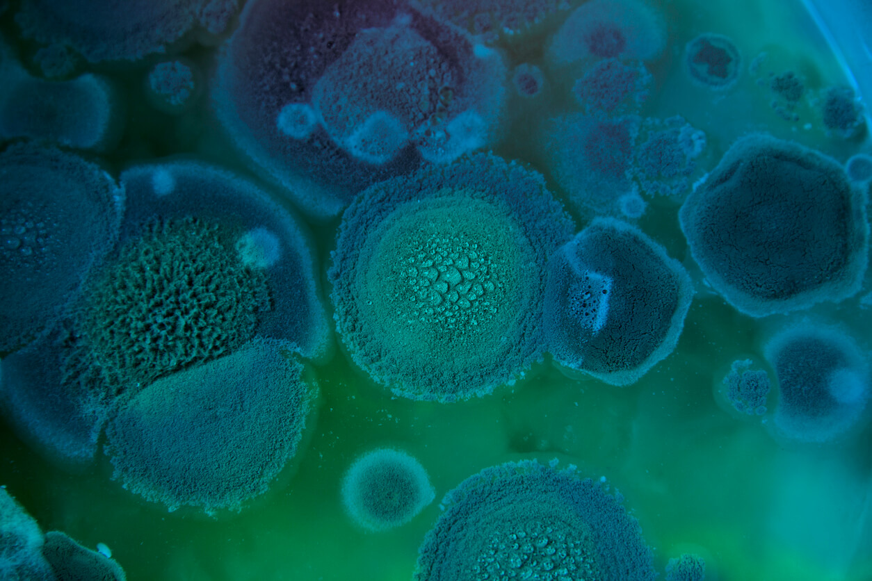
Parasites
Acanthamoeba
This is a microscopic, free-living ameba, or amoeba (single-celled living organism), that can cause rare, but severe infections of the eye, skin, and central nervous system. The ameba is found worldwide in the environment in water and soil. The ameba can be spread to the eyes through contact lens use, cuts, or skin wounds or by being inhaled into the lungs. Most people will be exposed to Acanthamoeba during their lifetime, but very few will become sick from this exposure.
Amebiasis
Amebiasis is a disease caused by the parasite Entamoeba histolytica. It can affect anyone, although it is more common in people who live in tropical areas with poor sanitary conditions. Diagnosis can be difficult because other parasites can look very similar to E. histolytica when seen under a microscope. Infected people do not always become sick. If your doctor determines that you are infected and need treatment, medication is available.
Angiostrongyliasis
This is a parasite nematode that can cause severe gastrointestinal or central nervous system disease in humans depending on the species. Angiostrongylis cantonensis, which is also known as the rat lung worm, causes eosinophilic meningitis and is prevalent in Southeast Asia and tropical Pacific islands. The distribution of this parasite has been increasing over time and infections have been identified in Africa, the Caribbean and the United States. Angiostrongylis costaricensis causes eosinophilic gastroenteritis and is found in Latin America and the Caribbean.
Anisakiasis
This is caused by anisakid nematodes (worms) that can invade the stomach wall or intestine of humans. The transmission of this disease occurs when infective larvae are ingested from fish or squid that humans eat raw or undercooked. In some cases, this infection is treated by removal of the larvae via endoscopy or surgery.
Babesiosis
Babesiosis is caused by microscopic parasites that infect red blood cells and are spread by certain ticks. Babesia microti is transmitted by the bite of infected Ixodes scapularis ticks-typically, by the nymph stage of the tick, which is about the size of a poppy seed. Although many people who are infected with Babesia do not have symptoms, for those who do effective treatment is available. Babesiosis is preventable, if simple steps are taken to reduce exposure to ticks.
Balantidiasis
Balantidium coli, though rare in the US, is an intestinal protozoan parasite that can infect humans. These parasites can be transmitted through the fecal-oral route by contaminated food and water.Balantidium coli infection is mostly asymptomatic, but people with other serious illnesses can experience persistent diarrhea, abdominal pain, and sometimes a perforated colon. When traveling to endemic tropical countries, Balantidium coli infection can be prevented by following good hygiene practices. Wash all fruits and vegetables with clean water when preparing or eating them, even if they have a removable skin.
Balamuthia
Balamuthia mandrillaris is a free-living ameba (a single-celled living organism) naturally found in the environment. Balamuthia can cause a rare and serious infection of the brain and spinal cord called Granulomatous Amebic Encephalitis (GAE).
Baylisascaris
This is a genus of roundworms. The microscopic larvae hatch in the intestine and invade the intestinal wall. If they are in their definitive host they develop for several weeks, then enter the intestinal lumen, mature, mate, and produce eggs, which are carried out in the fecal stream. If the larvae are in a paratenic host, they break into the bloodstream and enter various organs, particularly the central nervous system. The disease that causes it is Baylisascariasis. A great deal of damage occurs wherever the larva try to make a home. In response to the attack, the body attempts to destroy it by walling it off or killing it. The larva moves rapidly to escape, seeking out the liver, eyes, spinal cord or brain. Occasionally they can be found in the heart, lungs, and other organs. Eventually the larva dies and is reabsorbed by the body. In very small species such as mice, it might take only one or two larvae in the brain to be fatal. If the larva does not cause significant damage in vital organs, then the victim will show no signs of disease. On the other hand, if it causes behavioral changes by destroying parts of the brain, the host becomes easier prey, bringing the larva into the intestine of a new host.
Clinical signs in humans
Skin irritations from larvae migrating within the skin.
Respiratory discomfort, liver enlargement, and fever due to reaction to larvae migration.
Eye and brain tissue damage due to the random migration of the larvae.
Nausea, a lethargic feeling, incoordination and loss of eyesight.
Severe neurological signs including imbalance, circling and abnormal behavior, caused by extensive tissue damage due to larval migration through the brain, eventually seizures and coma. Source
Bed Bugs
Bed bugs, a problem worldwide, are resurging, causing property loss, expense, and inconvenience. The good news is that bed bugs do not transmit disease. The best way to prevent bed bugs is regular inspection for signs of an infestation.
Blastocystis Hominis
This is a common microscopic organism that inhabits the intestine and is found throughout the world. A full understanding of the biology of Blastocystis and its relationship to other organisms is not clear, but is an active area of research.
Capillariasis
Capillariasis is a parasitic disease in humans caused by two different species of capillarids: Capillaria hepatica and Capillaria philippinensis.C. hepatica is transferred through the fecal matter of infected animals and can lead to hepatitis. C. philippinensis is transferred through ingesting infected small freshwater fish and can lead to diarrhea and emaciation.
Cercarial Dermatitis
Swimmer’s itch, also called cercarial dermatitis, appears as a skin rash caused by an allergic reaction to certain parasites that infect some birds and mammals. These microscopic parasites are released from infected snails into fresh and salt water (such as lakes, ponds, and oceans). While the parasite’s preferred host is the specific bird or mammal, if the parasite comes into contact with a swimmer, it burrows into the skin causing an allergic reaction and rash. Swimmer’s itch is found throughout the world and is more frequent during summer months. Most cases of swimmer’s itch do not require medical attention.
Chagas Disease (AmericanTrypanosomiasis)
This is caused by the parasite Trypanosoma cruzi, which is transmitted to animals and people by insect vectors and is found only in the Americas (mainly, in rural areas of Latin America where poverty is widespread).
Chilomastix Mesnili
This is a non-parasitic member of primate gastrointestinal microflora, commonly associated with but not causing parasitic infections. It is found in about 3.5% of the population in the United States. In addition to humans, Chilomastix is found in chimpanzees, orangutans, monkeys, and pigs. It lives in the cecum and colon. C. mesnili has a similar life style to Giardia lamblia.
Although Chilomastix mesnili is considered non-pathogenic, it often occurs with other parasite infections. C. mesnili may be confused with other pathogenic species during diagnosis. It can create a false positive which would result in unnecessary treatment or a false negative which would withhold necessary treatment. Source
Clonorchiasis
This is a liver fluke that can infect the liver, gallbladder and bile duct in humans. Found across parts of Asia, it is also known as the Chinese or oriental liver fluke. Humans become infected when eating the parasite containing cysts within infected raw or undercooked fish, crabs, or crayfish. Clonorchiscan be treated by medication through your health professional.
CLM (Cutaneous Larva Migrans, Ancylostomiasis, Hookworm)
Zoonotic hookworms are hookworms that live in animals but can be transmitted to humans. Dogs and cats can become infected with several hookworm species, including Ancylostoma brazilense, A. caninum, A. ceylanicum, and Uncinaria stenocephala. The eggs of these parasites are shed in the feces of infected animals and can end up in the environment, contaminating the ground where the animal defecated. People become infected when the zoonotic hookworm larvae penetrate unprotected skin, especially when walking barefoot or sitting on contaminated soil or sand. This can result in a disease called cutaneous larva migrans (CLM), when the larvae migrate through the skin and cause inflammation.
Crabs
Pithariasis Adult pubic lice are 1.1-1.8 mm in length. Pubic lice typically are found attached to hair in the pubic area but sometimes are found on coarse hair elsewhere on the body (for example, eyebrows, eyelashes, beard, mustache, chest, armpits, etc.). Pubic lice infestations (pthiriasis) are usually spread through sexual contact. Dogs, cats, and other pets do not play a role in the transmission of human lice.
Cryptosporidiosis
Cryptosporidium is a microscopic parasite that causes the diarrheal disease cryptosporidiosis. Both the parasite and the disease are commonly known as “Crypto.”
There are many species of Cryptosporidium that infect animals, some of which also infect humans. The parasite is protected by an outer shell that allows it to survive outside the body for long periods of time and makes it very tolerant to chlorine disinfection.
While this parasite can be spread in several different ways, water (drinking water and recreational water) is the most common way to spread the parasite. Cryptosporidium is a leading cause of waterborne disease among humans in the United States.
Cyclosporiasis
Cyclosporiasis is an intestinal illness caused by the microscopic parasite Cyclospora cayetanensis. People can become infected withCyclospora by consuming food or water contaminated with the parasite. People living or traveling in countries where cyclosporiasis is endemic may be at increased risk for infection.
Cysticercosis
Cysticercosis is a parasitic tissue infection caused by larval cysts of the tapeworm Taenia solium. These larval cysts infect brain, muscle, or other tissue, and are a major cause of adult onset seizures in most low-income countries. A person gets cysticercosis by swallowing eggs found in the feces of a person who has an intestinal tapeworm. People living in the same household with someone who has a tapeworm have a much higher risk of getting cysticercosis than people who don’t. People do not get cysticercosis by eating undercooked pork. Eating undercooked pork can result in intestinal tapeworm if the pork contains larval cysts. Pigs become infected by eating tapeworm eggs in the feces of a human infected with a tapeworm. Both the tapeworm infection, also known as taeniasis, and cysticercosis occur globally. The highest rates of infection are found in areas of Latin America, Asia, and Africa that have poor sanitation and free-ranging pigs that have access to human feces.
Cystoisospora (formerly known as isosporiasis)
This is an intestinal disease of humans caused by the coccidian parasite Cystoisospora belli (formerly known asIsospora belli). Cystoisosporiasis is most common in tropical and subtropical areas of the world. The parasite can be spread by ingesting contaminated food or water. The most common symptom is watery diarrhea. The infection is treatable and preventable.
Dientamoeba Fragilis
This is a parasite that lives in the large intestine of people. This protozoan parasite produces trophozoites; cysts have not been identified. The intestinal infection may be either asymptomatic or symptomatic.
Diphyllobothriasis
Diphyllobothrium latum and related species (the fish or broad tapeworm), the largest tapeworms that can infect people, can grow up to 30 feet long. While most infections are asymptomatic, complications include intestinal obstruction and gall bladder disease caused by migration of proglottids. Diagnosis is made by identification of eggs or segments of the tapeworm in a stool sample with a microscope. Safe and effective medications are available to treat Diphyllobothrium. Infections are acquired by eating raw or undercooked fish, usually from the Northern Hemisphere (Europe, newly independent states of the Former Soviet Union, North America, Asia), but cases have also been reported in Uganda and Chile.
Dipylidium
This is tapeworm of cats and dogs. People become infected when they accidentally swallow a flea infected with tapeworm larvae; most reported cases involve children. Dipylidium infection is easily treated in humans and animals.
Dirofilariasis
Dirofilariasis in humans is caused by Dirofilaria roundworms. The main natural hosts for the three Dirofilaria species that most frequently cause disease in humans are dogs and wild canids (such as wolves and foxes) and raccoons. Humans are infected with Dirofilaria larvae through mosquito bites. Infection can result in nodules under the skin or conjunctiva and lung granulomas (small nodules formed by an inflammatory reaction) that appear as coin lesions (small, round abnormalities) on x-rays, leading to diagnostic procedures to exclude more serious diseases.
Dracunculiasis (Guinea Worm Disease)
This is considered a neglected tropical disease, is caused by the parasite Dracunculus medinensis. The disease affects poor communities in remote parts of Africa that do not have safe water to drink. There is no drug treatment for Guinea worm disease nor a vaccine to prevent it.
Echinococcosis (Hytadid)
This is a parasitic disease caused by infection with tiny tapeworms of the genus Echinocococcus. There are two types Cystic echinocccosis (CE), is caused by infection with the larval stage of Echinococcus granulosus, a ~2-7 millimeter long tapeworm found in dogs (definitive host) and sheep, cattle, goats, and pigs (intermediate hosts). Although most infections in humans are asymptomatic, CE causes harmful, slowly enlarging cysts in the liver, lungs, and other organs that often grow unnoticed and neglected for years. Alveolar echinococcosis (AE) disease is caused by infection with the larval stage of Echinococcus multilocularis, a ~1-4 millimeter long tapeworm found in foxes, coyotes, and dogs (definitive hosts). Small rodents are intermediate hosts for E. multilocularis. Although cases of AE in animals in endemic areas are relatively common, human cases are rare. AE poses a much greater health threat to people than CE, causing parasitic tumors that can form in the liver, lungs, brain, and other organs. If left untreated, AE can be fatal.
Elephantiasis
This is a symptom of a variety of diseases, where parts of a person’s body swell to massive proportions.
Some conditions that have this symptom include:
Elephantiasis nostras, due to longstanding chronic lymphangitis
Elephantiasis tropica or lymphatic filariasis, caused by a number of parasitic worms, particularly Wuchereria bancrofti. More than 120 million people, mostly in Africa and Southeast Asia, are affected.
Nonfilarial elephantiasis or podoconiosis, an immune disease affecting the lymph vessels
Elephantiasis, Grade 3 lymphedema which may occur in people with breast cancer.
Genital elephantiasis, end result of lymphogranuloma venereum
Proteus syndrome, the genetic disorder of the so-called Elephant Man. Source
Endolimax Nana
This is the smallest of the intestine-dwelling amoebae infecting humans, its trophozoite averaging only 8 μm in diameter (range, 6-15 μm). The trophozoite lives in the host’s colon and is generally considered to be nonpathogenic. According to some surveys, prevalence may be as high as 30% in some populations. The life cycle is identical to that of other cyst-forming amoebae, with the cyst being the infective stage. E. nana cysts can be identified and distinguished from other cysts by their smaller size (9 μm in greatest diameter; range, 5-14 μm, ovoid shape, and one to four vesicular nuclei, each usually containing a large, eccentric endosome). Source
Entamoeba Coli
This is a non-pathogenic species of Entamoeba that frequently exists as a commensal parasite in the human gastrointestinal tract. E. coli (not to be confused with the bacterium Escherichia coli) is important in medicine because it can be confused during microscopic examination of stained stool specimens with the pathogenic Entamoeba histolytica. This amoeba does not move much by the use of its pseudopod, and creates a “sur place (non-progressive) movement” inside the large intestine. Usually, the amoeba is immobile, and keeps its round shape. This amoeba, in its trophozoite stage, is only visible in fresh, unfixed stool specimens. Sometimes the Entamoeba coli have parasites as well. One is the fungus Sphaeritaspp. This fungus lives in the cytoplasm of the E. coli. While this differentiation is typically done by visual examination of the parasitic cysts via light microscopy, new methods using molecular biology techniques have been developed. The scientific name of the amoeba, E. coli, is often mistaken for the bacterium, Escherichia coli. Unlike the bacterium, the amoeba is mostly harmless, and does not cause as many intestinal problems as some strains of the E. coli bacterium. Some of these harmful strains are inside raw or uncooked meat that is consumed. For example, the bacterium, E. coli O157:H7, which can cause illness, and even death, if eaten. To make the naming of these organisms less confusing, “alternate contractions” are used to name the species for the purpose making the naming easier; for example, using Esch. coli and Ent. coli for the bacterium and amoeba, instead of using E. coli for both. Source
Entamoeba Dispar
Entamoeba Hartmanni
Entamoeba Histolytica
Amebiasis is a disease caused by the parasite Entamoeba histolytica. It can affect anyone, although it is more common in people who live in tropical areas with poor sanitary conditions. Diagnosis can be difficult because other parasites can look very similar to E. histolytica when seen under a microscope. Infected people do not always become sick. If your doctor determines that you are infected and need treatment, medication is available.
Entamoeba Polecki
Entamoeba polecki is a single-celled parasite that is found in intestines, mainly in pigs and monkeys. Other animals that it can be found in are cattle, goats, sheep, dogs, and humans. The way humans get infected is by swallowing the parasite. Source
Fascioliasis
Fascioliasis is a parasitic infection typically caused by Fasciola hepatica, which is also known as “the common liver fluke” or “the sheep liver fluke.” A related parasite, Fasciola gigantica, also can infect people. Fascioliasis is found in all 5 continents, in over 50 countries, especially where sheep or cattle are reared. People usually become infected by eating raw watercress or other water plants contaminated with immature parasite larvae. The immature larval flukes migrate through the intestinal wall, the abdominal cavity, and the liver tissue, into the bile ducts, where they develop into mature adult flukes, which produce eggs. The pathology typically is most pronounced in the bile ducts and liver. Fasciola infection is both treatable and preventable.
Fasciolopsiasis
The intestinal fluke Fasciolopsis buski, which causes faciolopsiasis, is the largest intestinal fluke of humans. Fasciolopsiasis can be prevented by cooking aquatic plants well before eating them. Fasciolopsis is found in south and southeastern Asia. Fasciolopsiasis is treatable.
Giardiasis
Giardia is a microscopic parasite that causes the diarrheal illness known as giardiasis. Giardia (also known as Giardia intestinalis, Giardia lamblia, or Giardia duodenalis) is found on surfaces or in soil, food, or water that has been contaminated with feces (poop) from infected humans or animals.
Giardia is protected by an outer shell that allows it to survive outside the body for long periods of time and makes it tolerant to chlorine disinfection. While the parasite can be spread in different ways, water (drinking water and recreational water) is the most common mode of transmission.
Gnathostomiasis
Human gnathostomiasis is caused by several species of parasitic worms (nematodes) in the genus Gnathostoma. The disease is found and is most commonly diagnosed in Southeast Asia, though it has also been found elsewhere in Asia, in South and Central America, and in some areas of Africa. People become infected primarily by eating undercooked or raw freshwater fish, eels, frogs, birds, and reptiles. The most common manifestations of the infection in humans are migratory swellings under the skin and increased levels of eosinophils in the blood. Rarely, the parasite can enter other tissues such as the liver, and the eye, resulting in vision loss or blindness, and the nerves, spinal cord, or brain, resulting in nerve pain, paralysis, coma and death.
Heterophyiasis
The definitive host becomes infected by ingesting undercooked or salted fish containing metacercariae. After ingestion, the metacercariae excyst, attach to the mucosa of the small intestine and mature into adults. The main symptoms are diarrhea and colicky abdominal pain. Migration of the eggs to the heart, resulting in potentially fatal myocardial and valvular damage, has been reported from the Philippines. Migration to other organs (e.g., brain) has also been reported.
Hookworm Human
Hookworm, Ascaris, and whipworm are known as soil transmitted helminths (parasitic worms). Together, they account for a major burden of disease worldwide. Hookworms live in the small intestine. Hookworm eggs are passed in the feces of an infected person. If the infected person defecates outside (near bushes, in a garden, or field) of if the feces of an infected person are used as fertilizer, eggs are deposited on soil. They can then mature and hatch, releasing larvae (immature worms). The larvae mature into a form that can penetrate the skin of humans. Hookworm infection is mainly acquired by walking barefoot on contaminated soil. One kind of hookworm can also be transmitted through the ingestion of larvae.
Most people infected with hookworms have no symptoms. Some have gastrointestinal symptoms, especially persons who are infected for the first time. The most serious effects of hookworm infection are blood loss leading to anemia, in addition to protein loss. Hookworm infections are treatable with medication prescribed by your health care provider.
Hookworm Zoonotic
Zoonotic hookworms are hookworms that live in animals but can be transmitted to humans. Dogs and cats can become infected with several hookworm species, including Ancylostoma brazilense, A. caninum, A. ceylanicum, and Uncinaria stenocephala. The eggs of these parasites are shed in the feces of infected animals and can end up in the environment, contaminating the ground where the animal defecated. People become infected when the zoonotic hookworm larvae penetrate unprotected skin, especially when walking barefoot or sitting on contaminated soil or sand. This can result in a disease called cutaneous larva migrans (CLM), when the larvae migrate through the skin and cause inflammation.
Hymenolepiasis
This infection with the dwarf tapeworm is found worldwide. It is most often seen in children in countries in which sanitation and hygiene are inadequate. Although the dwarf tapeworm infection rarely causes symptoms, it can be misdiagnosed for pinworm infection.
Kala-azar
Leishmaniasis is a parasitic disease that is found in parts of the tropics, subtropics, and southern Europe. It is classified as a neglected tropical disease. Leishmaniasis is caused by infection with Leishmania parasites, which are spread by the bite of phlebotomine sand flies. There are several different forms of leishmaniasis in people. The most common forms are cutaneous leishmaniasis, which causes skin sores, and visceral leishmaniasis, which affects several internal organs (usually spleen, liver, and bone marrow).
Keratitis
This is a condition in which the eye‘s cornea, the clear dome on the front surface of the eye, becomes inflamed. The condition is often marked by moderate to intense pain and usually involves any of the following symptoms: pain, impaired eyesight, photophobia (light sensitivity), red eye and a ‘gritty’ sensation. Source
Loiasis
Loiasis, called African eye worm by most people, is caused by the parasitic worm Loa loa. It is passed on to humans through the repeated bites of deerflies (also known as mango flies or mangrove flies) of the genus Chrysops. The flies that pass on the parasite breed in certain rain forests of West and Central Africa. Infection with the parasite can also cause repeated episodes of itchy swellings of the body known as Calabar swellings. Knowing whether someone has a Loa loa infection has become more important in Africa because the presence of people with Loa loa infection has limited programs to control or eliminate (river blindness) and (elephantiasis). There may be more than 29 million people who are at risk of getting loaisis in affected areas of Central and West Africa.
Lymph Filariasis
Lymphatic filariasis, considered globally as a neglected tropical disease, is a parasitic disease caused by microscopic, thread-like worms. The adult worms only live in the human lymph system. The lymph system maintains the body’s fluid balance and fights infections. Lymphatic filariasis is spread from person to person by mosquitoes. People with the disease can suffer from lymphedema and elephantiasis and in men, swelling of the scrotum, called hydrocele. Lymphatic filariasis is a leading cause of permanent disability worldwide. Communities frequently shun and reject women and men disfigured by the disease. Affected people frequently are unable to work because of their disability, and this harms their families and their communities.
Malaria
This is a mosquito-borne disease caused by a parasite. People with malaria often experience fever, chills, and flu-like illness. Left untreated, they may develop severe complications and die.
Microsporidiosis
The microsporidia are a group of obligate intracellular parasitic fungi. Historically, they have been treated among the protozoa, and as such are often still managed by diagnostic parasitology laboratories. To date, more than 1,200 species belonging to 143 genera have been described as parasites infecting a wide range of vertebrate and invertebrate hosts. Microsporidia, are characterized by the production of resistant spores that vary in size, depending on the species. They possess a unique organelle, the polar tubule or polar filament, which is coiled inside the spore as demonstrated by its ultrastructure. The microsporidia spores of species associated with human infection measure from 1 to 4 µm and that is a useful diagnostic feature. There are at least 15 microsporidian species that have been identified as human pathogens: Anncaliia (formerly Brachiola) algerae, A. connori, A. vesicularum, Encephalitozoon cuniculi, E. hellem, E. intestinalis, Enterocytozoon bieneusi Microsporidium ceylonensis, M. africanum, Nosema ocularum, Pleistophora sp.,Trachipleistophora hominis, T. anthropophthera, Vittaforma corneae, and Tubulinosema acridophagus. Encephalitozoon intestinalis
Myiasis
Myiasis is the infection of a fly larva (maggot) in human tissue. This occurs in tropical and subtropical areas. Myiasis is rarely acquired in the United States; people typically get the infection when they travel to tropical areas in Africa and South America. People traveling with untreated and open wounds are more at risk for getting myiasis. Fly larvae need to be surgically removed by a medical professional.
Naegleria
Naegleria fowleri (commonly referred to as the “brain-eating amoeba” or “brain-eating ameba”), is a free-living microscopic ameba, (single-celled living organism). It can cause a rare and devastating infection of the brain called primary amebic meningoencephalitis (PAM). The ameba is commonly found in warm freshwater (e.g. lakes, rivers, and hot springs) and soil. Naegleria fowleri usually infects people when contaminated water enters the body through the nose. Once the ameba enters the nose, it travels to the brain where it causes PAM, which is usually fatal. Infection typically occurs when people go swimming or diving in warm freshwater places, like lakes and rivers. In very rare instances, Naegleria infections may also occur when contaminated water from other sources (such as inadequately chlorinated swimming pool water or heated and contaminated tap water) enters the nose. You cannot get infected from swallowing water contaminated with Naegleria.
Neurocysticercosis
Cysticercosis is a parasitic infection that results from ingestion of eggs from the adult tapeworm, Taenia solium (T. solium) (1,2). When cysticercosis involves the central nervous system, it is called neurocysticercosis. Neurocysticercosis is the most common parasitic infection of the brain and a leading cause of epilepsy in the developing world, especially Latin America, India, Africa, and China (1–12). Source
Opisthorchiasis
Opisthorchis species are liver fluke parasites acquired by eating raw or undercooked fish from endemic countries: Thailand, Laos, Cambodia, Viet Nam, and other areas of Asia, Eastern Europe, and the former Soviet Union. While most infected persons are asymptomatic, infections of longer duration can result in severe symptoms and serious illness. Diagnosis is based on identification of eggs in stool specimens with a microscope. Safe and effective medication is available to treat Opisthorchis infections. Adequately freezing or cooking fish will kill the parasite.
Paragonimiasis
Paragonimus is a lung fluke (flatworm) that infects the lungs of humans after eating an infected raw or undercooked crab or crayfish. Less frequent, but more serious cases of paragonimiasis occur when the parasite travels to the central nervous system. Although rare, paragonimiasis has been acquired in the United States, with multiple cases reported from the Midwest. Once the diagnosis is made, effective treatment for paragonimiasis is available from a physician.
Pediculosis Lice
Lice are parasitic insects that can be found on people’s heads, and bodies, including the pubic area. Human lice survive by feeding on human blood. Lice found on each area of the body are different from each other. Pediculus humanus capitis (head louse) and Pediculus humanus corporis(body louse, clothes louse). Only the body louse is known to spread disease. Lice infestations are spread most commonly by close person-to-person contact. Dogs, cats, and other pets do not play a role in the transmission of human lice. Lice move by crawling; they cannot hop or fly. Both over-the-counter and prescription medications are available for treatment of lice infestations.
Pneumocystis
Pneumocystis jirovecii (previously classified as Pneumocystis carinii) was previously classified as a protozoa. Currently, it is considered a fungus based on nucleic acid and biochemical analysis.
River Blindness (Onchocerciasis)
This is a neglected tropical disease caused by the parasitic worm Onchocerca volvulus. It is transmitted through repeated bites by blackflies of the genus Simulium. The disease is called River Blindness because the blackfly that transmits the infection lives and breeds near fast-flowing streams and rivers and the infection can result in blindness. In addition to visual impairment or blindness, onchocerciasis causes skin disease, including nodules under the skin or debilitating itching. Worldwide onchocerciasis is second only to trachoma as an infectious cause of blindness.
Roundworm Human (ascariasis)
Ascaris lives in the intestine and Ascaris eggs are passed in the feces of infected persons. If the infected person defecates outside (near bushes, in a garden, or field) or if the feces of an infected person are used as fertilizer, eggs are deposited on soil. They can then mature into a form that is infective. Ascariasis is caused by ingesting eggs. This can happen when hands or fingers that have contaminated dirt on them are put in the mouth or by consuming vegetables or fruits that have not been carefully cooked, washed or peeled. People infected with Ascaris often show no symptoms. If symptoms do occur they can be light and include abdominal discomfort. Heavy infections can cause intestinal blockage and impair growth in children. Other symptoms such as cough are due to migration of the worms through the body. Ascariasis is treatable with medication prescribed by your health care provider.
Roundworm Raccoon
Baylisascaris infection is caused by a roundworm found in raccoons. This roundworm can infect people as well as a variety of other animals, including dogs. Human infections are rare, but can be severe if the parasites invade the eye (ocular larva migrans), organs (visceral larva migrans) or the brain (neural larva migrans).
Sappinia
This is a free-living ameba, or amoeba (a single-celled living organism), found in the environment. There are two known species of Sappinia: Sappinia diploidea and Sappinia pedata. This ameba causes amebic encephalitis, which is an infection of the brain. Worldwide, only one case of amebic encephalitis due to Sappinia infection has been reported.
Sarcocystosis
Sarcocystosis is a disease caused by a microscopic parasite Sarcocystis. In humans, two types of the disease can occur, one causes diarrhea, mild fever, and vomiting (intestinal type), and the other type causes muscle pain, transitory edema, and fever (muscular type). However, most people infected with Sarcocystis do not have symptoms. Sarcocystosis occurs in tropical or subtropical countries. Muscular sarcocystosis has most often been reported from countries in Southeast Asia.
Scabies
Human scabies is caused by an infestation of the skin by the human itch mite (Sarcoptes scabiei var. hominis). The microscopic scabies mite burrows into the upper layer of the skin where it lives and lays its eggs. The most common symptoms of scabies are intense itching and a pimple-like skin rash. The scabies mite usually is spread by direct, prolonged, skin-to-skin contact with a person who has scabies.
Scabies occurs worldwide and affects people of all races and social classes. Scabies can spread rapidly under crowded conditions where close body contact is frequent. Institutions such as nursing homes, extended-care facilities, and prisons are often sites of scabies outbreaks.
Schistosomiasis
Schistosomiasis, also known as bilharzia, is a disease caused by parasitic worms. Although the worms that cause schistosomiasis are not found in the United States, more than 200 million people are infected worldwide. In terms of impact this disease is second only to malaria as the most devastating parasitic disease. The parasites that cause schistosomiasis live in certain types of freshwater snails. The infectious form of the parasite, known as cercariae, emerge from the snail, hence contaminating water. You can become infected when your skin comes in contact with contaminated freshwater. Most human infections are caused by Schistosoma mansoni, S. haematobium, or S. japonicum.
Strongyloidiasis
It is a parasitic disease caused by nematodes, or roundworms, in the genus Strongyloides that enter the body through exposed skin, such as bare feet. Strongyloides is most common in tropical or subtropical climates.Most people who are infected with Strongyloides do not know they are infected and have no symptoms. Others may develop a severe form and, if untreated, become critically ill and potentially die.
Tapeworm (Human)
Taeniasis in humans is a parasitic infection caused by the tapeworm species Taenia saginata (beef tapeworm), Taenia solium (pork tapeworm), and Taenia asiatica (Asian tapeworm). Humans can become infected with these tapeworms by eating raw or undercooked beef (T. saginata) or pork (T. soliumand T. asiatica). People with taeniasis may not know they have a tapeworm infection because symptoms are usually mild or nonexistent. Taenia soliumtapeworm infections can lead to cysticercosis, which is a disease that can cause seizures, so it is important seek treatment.
Toxocariasis (Roundworm)
This can also cause Ocular Larva Migrans: Toxocariasis is the parasitic disease caused by the larvae of two species of Toxocararoundworms: Toxocara canis from dogs and, less commonly, Toxocara cati from cats. Toxocariasis is considered one of the neglected parasitic infectionshttps://www.cdc.gov/parasites/npi, a group of five parasitic diseases that have been targeted by CDC for public health action.
Toxoplasmosis
Toxoplasmosis is considered to be a leading cause of death attributed to foodborne illness in the United States. More than 60 million men, women, and children in the U.S. carry the Toxoplasma parasite, but very few have symptoms because the immune system usually keeps the parasite from causing illness. However, women newly infected with Toxoplasma during pregnancy and anyone with a compromised immune system should be aware that toxoplasmosis can have severe consequences.
Trichinellosis
Trichinellosis, also called trichinosis, is a disease that people can get by eating raw or undercooked meat from animals infected with the microscopic parasite Trichinella.
Trichomoniasis
This is a sexually transmitted disease (STD). Both men and women can get trichomoniasis. Many people who have trichomoniasis don’t know it. The infection often has no symptoms. Women are more likely than men to get symptoms. You can pass trichomoniasis to others without knowing it. Trichomoniasis is easy to treat and cure. Women should be tested for trichomoniasis if you have any of these symptoms: A discharge from your vagina, pain when you have sex, pain when you pass urine.
Trichuriasis
An estimated 604-795 million people in the world are infected with whipworm. Whipworm, hookworm, and Ascaris are known as soil transmitted helminths (parasitic worms). Together, they account for a major burden of disease worldwide. Whipworms live in the large intestine and whipworm eggs are passed in the feces of infected persons. If the infected person defecates outside (near bushes, in a garden, or field) or if human feces as used as fertilizer, eggs are deposited on soil. They can then mature into a form that is infective. Whipworm infection is caused by ingesting eggs. This can happen when hands or fingers that have contaminated dirt on them are put in the mouth.
Trypanosomiasis
This is the name of several diseases in vertebrates caused by parasiticprotozoan trypanosomes of the genus Trypanosoma. In humans this includes African trypanosomiasisand Chagas disease.
The tsetse fly bite erupts into a red chancre sore and within a few weeks, the person can experience fever, swollen lymph glands, blood in urine, aching muscles and joints, headaches and irritability. In the first phase, the patient has only intermittent bouts of fever with lymphadenopathy together with other non-specific signs and symptoms. The second stage of the disease is marked by involvement of the central nervous system with extensive neurological effects like changes in personality, alteration of the biological clock (the circadian rhythm), confusion, slurred speech, seizures and difficulty in walking and talking. These problems can develop over many years and if not treated, the person dies. It is common to the African continent. Source
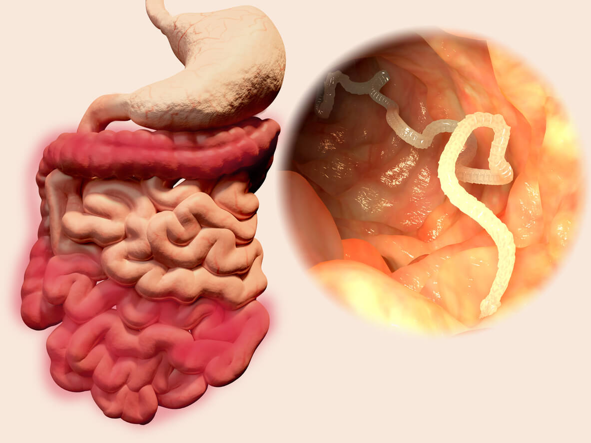
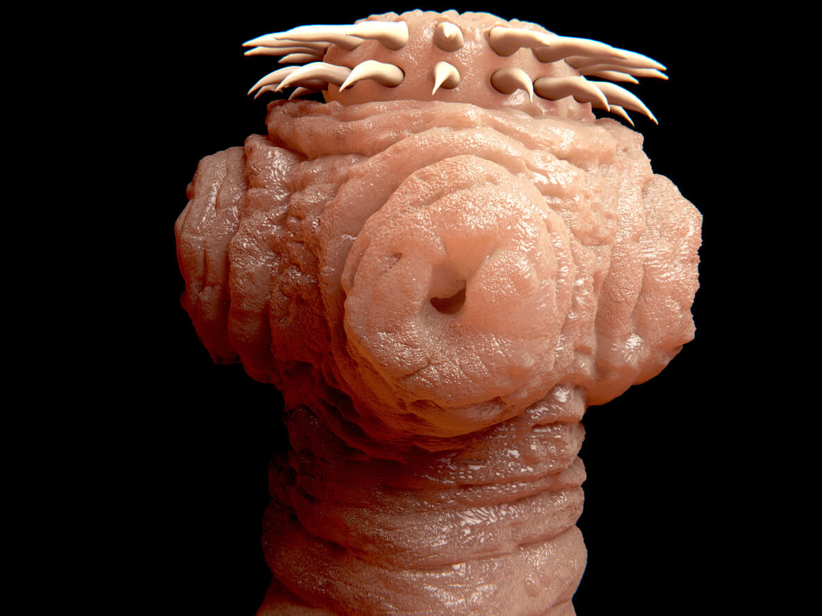
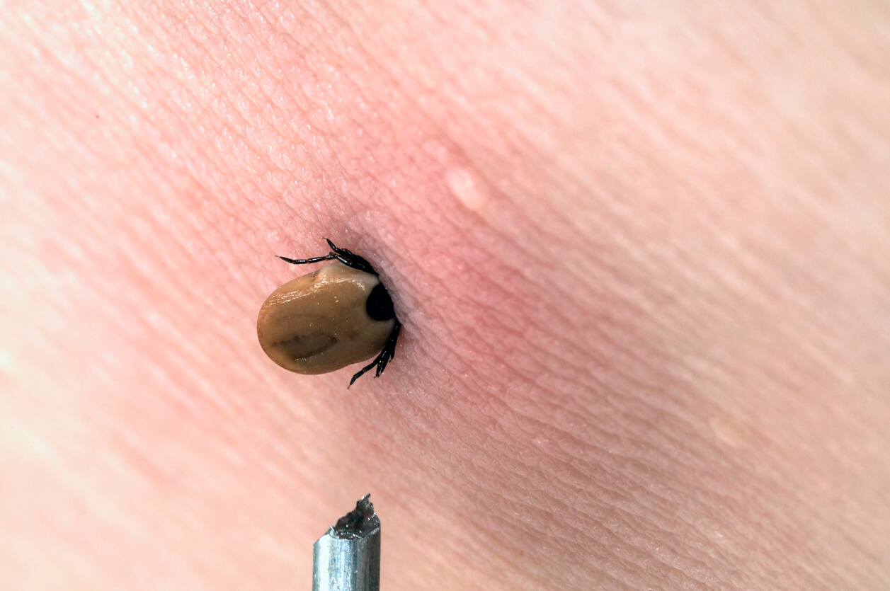
Viruses
Bornavirus (BDV)
Coxsackie Virus
Cytomegalovirus (CMV)
Signs and Symptoms
CMV usually causes an asymptomatic infection or produces mild flulike symptoms; afterward, it remains latent throughout life and may reactivate. Most patients with CMV infection exhibit few clinical findings on physical examination. Primary CMV infection may be a cause of fever of unknown origin. Symptoms, when apparent, develop 9-60 days after primary infection.
Pharyngitis may be present.
Examination of the lungs may reveal fine crackles.
The lymph nodes and spleen may be enlarged, so CMV should be included in the differential diagnoses of infections that produce lymphadenopathy.
In immunocompromised individuals, symptomatic disease usually manifests as a mononucleosis syndrome. Symptomatic CMV disease can affect almost every organ of the body, resulting in fever of unknown origin, pneumonia, hepatitis, encephalitis, myelitis, colitis, uveitis, retinitis, and neuropathy. Rarer manifestations of CMV infections in immunocompetent individuals include Guillain-Barré syndrome, meningoencephalitis, pericarditis, myocarditis, thrombocytopenia, and hemolytic anemia.
In patients with HIV infection, CMV involves the entire GI tract. Retinitis is the most common manifestation of CMV disease in patients who are HIV positive.
Epstein Barr Virus (EBV)
Herpes Simplex 1(HSV-1)
HSV-1 is the main cause of herpes infections on the mouth and lips, including cold sores and fever blisters. It is transmitted through kissing or sharing drinking glasses and utensils. HSV-1 can also cause genital herpes, although HSV-2 is the main cause of genital herpes. Exposure to HSV-1 is extremely common, as many as 90% of American adults have been exposed to the virus, and there is no stigma to having a cold sore.
Symptoms may include:
Small, painful, fluid-filled blisters around the lips or edge of the mouth
Tingling or burning around the mouth or nose, often a few days before blisters appear
Fever
Sore throat
Swollen lymph nodes in neck
Herpes Simplex 2 (HSV-2)
A herpes virus that causes genital herpes, characterized by sores in the genital area. It is a sexually transmitted disease. HSV-2 can also cause infection of the brain (encephalitis) if the immune system is severely defective or compromised. The treatment of infection with herpes simplex 2 is usually by topical or oral anti-viral medication, although intravenous therapy is required to treat infections of the brain (encephalitis).
Symptoms may include:
Tingling sensation in the genitalia, buttocks, and thighs
Small red blisters or open sores on genitals or inner thighs; in women, often occur inside the vagina
May be painful or not
In women, vaginal discharge
Fever, muscle aches
Headache
Painful urination
Swollen lymph glands in the groin
Hepatitis A
Hepatitis A (formerly known as infectious hepatitis) is an infectious disease of the liver caused by the hepatitis A virus (HAV). Many cases have few or no symptoms, especially in the young. The time between infection and symptoms, in those who develop them, is between two and six weeks. When symptoms occur, they typically last eight weeks and may include nausea, vomiting, diarrhea, jaundice, fever, and abdominal pain. Around 10-15% of people experience a recurrence of symptoms during the six months after the initial infection. Acute liver failure may rarely occur, with this being more common in the elderly. It is usually spread by eating food or drinking water contaminated with infected feces. Shellfish which have not been sufficiently cooked are a relatively common source. It may also be spread through close contact with an infectious person. While children often do not have symptoms when infected, they are still able to infect others. After a single infection, a person is immune for the rest of his or her life.
Hepatitis B
Hepatitis B is an infectious disease caused by the hepatitis B virus (HBV) that affects the liver. It can cause both acute and chronic infections. Many people have no symptoms during the initial infection. Some develop a rapid onset of sickness with vomiting, yellowish skin, tiredness, dark urine and abdominal pain. Often these symptoms last a few weeks and rarely does the initial infection result in death. It may take 30 to 180 days for symptoms to begin. In those who get infected around the time of birth 90% develop chronic hepatitis B while less than 10% of those infected after the age of five do. Most of those with chronic disease have no symptoms; however, cirrhosis and liver cancer may eventually develop. These complications result in the death of 15 to 25% of those with chronic disease.
Hepatitis C
The virus is transmitted by exposure to infectious blood or body fluids. Infection around the time of birth or from contact with other people’s blood during childhood is the most frequent method by which hepatitis B is acquired in areas where the disease is common. In areas where the disease is rare, intravenous drug use and sexual intercourse are the most frequent routes of infection. Other risk factors include working in healthcare, blood transfusions, dialysis, living with an infected person, travel in countries where the infection rate is high, and living in an institution. The hepatitis B viruses cannot be spread by holding hands, sharing eating utensils, kissing, hugging, coughing, sneezing, or breastfeeding.
Hepatitis C is an infectious disease caused by the hepatitis C virus (HCV) that primarily affects the liver. During the initial infection people often have mild or no symptoms. Occasionally a fever, dark urine, abdominal pain, and yellow tinged skin occurs. The virus persists in the liver in about 75% to 85% of those initially infected. Early on chronic infection typically has no symptoms. Over many years however, it often leads to liver disease and occasionally cirrhosis. In some cases, those with cirrhosis will develop complications such as liver failure, liver cancer, or esophageal and gastric varices.
HCV is spread primarily by blood-to-blood contact associated with intravenous drug use, poorly sterilized medical equipment, needlestick injuries in healthcare, and transfusions. Using blood screening, the risk from a transfusion is less than one per two million. It may also be spread from an infected mother to her baby during birth. It is not spread by superficial contact. There is no vaccine against hepatitis C.
Hepatitis D
Hepatitis D (hepatitis delta) is a disease caused by the hepatitis D virus (HDV), a small spherical enveloped viroid. HDV is considered to be a subviral satellite because it can propagate only in the presence of the hepatitis B virus (HBV). Transmission of HDV can occur either via simultaneous infection with HBV (coinfection) or superimposed on chronic hepatitis B or hepatitis B carrier state (superinfection).
Both superinfection and coinfection with HDV results in more severe complications compared to infection with HBV alone. These complications include a greater likelihood of experiencing liver failure in acute infections and a rapid progression to liver cirrhosis, with an increased risk of developing liver cancer in chronic infections. In combination with hepatitis B virus, hepatitis D has the highest fatality rate of all the hepatitis infections, at 20%.
Hepatitis E
Hepatitis E is a viral hepatitis (liver inflammation) caused by infection with a virus called hepatitis E virus.[1] It is one of five known human hepatitis viruses: A, B, C, D, and E. HEV is a positive-sense single-stranded non-enveloped RNA icosahedral virus, HEV has a fecal-oral transmission route.
Although Hepatitis E often causes an acute and self-limiting infection (the virus usually resolves itself and the individual recovers) with low mortality rates in the western world, it bears a high risk of developing chronic hepatitis in immunocompromised patients with substantial mortality rates. Organ transplant recipients who receive immunosuppressive medication to prevent rejection are thought to be the main population at risk for chronic hepatitis E. Furthermore, in healthy individuals during the duration of the infection, the disease severely impairs a person’s ability to work, care for family members, and other daily activities. Hepatitis E occasionally develops into an acute, severe liver disease, and is fatal in about 2% of all cases. Clinically, it is comparable to hepatitis A, but in pregnant women the disease is more often severe and is associated with a clinical syndrome called fulminant liver failure. Pregnant women, especially those in the third trimester, suffer an elevated mortality rate from the disease of around 20%.
Human Herpes Virus-6 (HHV-6)
This is the virus that most commonly causes the childhood disease, roseola. It was first discovered in 1986. Studies show that HHV-6 infects approximately 90% of children by age 2 years. It is usually marked by several days of high fever followed by a distinctive rash just as the fever breaks. In less than 1 percent of all adults, the virus can also slyly work its own DNA into the human genome. This makes it possible for mothers and fathers to pass HHV-6 to their children if these insertions are present in their eggs or sperm.
Human herpesvirus 6A (HHV-6A), A is rare, and acquired in adulthood, and human herpesvirus 6B (HHV-6B), B is common, usually acquired in childhood. Both A and B can reactivate at a later date, and are believed to contribute to diseases of the bone marrow and/or central nervous system in some people. HHV-6B has been associated with a variety of viral illnesses, including exanthem subitum, roseola infantum, fatal encephalitis, focal encephalitis, mononucleosis, lymphadenopathy, myocarditis, myelosuppression, and pneumonitis.
Many cases of HHV-6 infections are silent or appear with a fever, but HHV-6 infection in infants is the most common cause of fever-induced seizures usually associated with the primary HHV-6 infection. HHV-6 infection in adults is seen usually in those having a compromised immune system, those who have undergone organ transplants or in those with HIV infection.
New research suggests that HHV-6 may play a role in several chronic neurological conditions including MS (multiple sclerosis), mesial temporal lobe epilepsy, status epilepticus, fibromyalgia, and chronic fatigue syndrome.
Influenza A Virus
causes influenza in birds and some mammals, and is the only species of influenza virus A. Influenza virus A is a genus of the Orthomyxoviridae family of viruses. Strains of all subtypes of influenza A virus have been isolated from wild birds, although disease is uncommon. Some isolates of influenza A virus cause severe disease both in domestic poultry and, rarely, in humans. Occasionally, viruses are transmitted from wild aquatic birds to domestic poultry, and this may cause an outbreak or give rise to human influenza pandemics.
Influenza A viruses are negative-sense, single-stranded, segmented RNA viruses. The several subtypes are labeled according to an H number (for the type of hemagglutinin) and an N number (for the type of neuraminidase). There are 18 different known H antigens (H1 to H18) and 11 different known N antigens (N1 to N11). H17 was isolated from fruit bats in 2012. H18N11 was discovered in a Peruvian bat in 2013.
Each virus subtype has mutated into a variety of strains with differing pathogenic profiles; some are pathogenic to one species but not others, some are pathogenic to multiple species.
Influenza B
Influenzavirus B is a genus in the virus family Orthomyxoviridae. The only species in this genus is called Influenza B virus. Influenza B viruses are only known to infect humans and seals, giving them influenza. This limited host and range is apparently responsible for the lack of Influenzavirus B-caused influenza pandemics in contrast with those caused by the morphologically similar Influenzavirus A as both mutate by both antigenic drift and reassortment.
Currently there are two co-circulating lineages of the Influenza B virus based on the antigenic properties of the surface glycoprotein hemagglutinin. The lineages are termed B/Yamagata/16/88-like and B/Victoria/2/87-like viruses. The quadrivalent influenza vaccine licensed by the CDC is currently designed to protect against both co-circulating lineages and has been shown to have greater effectiveness in prevention of influenza caused by influenza B virus than the previous trivalent vaccine.
Further diminishing the impact of this virus “in man, influenza B viruses evolve slower than A viruses and faster than C viruses”. Influenzavirus B mutates at a rate 2 to 3 times slower than type A. It is currently accepted that influenza B viruses cause significant morbidity and mortality worldwide, and significantly impacts adolescents and schoolchildren.
Influenza C
Influenza viruses are members of the family Orthomyxoviridae. Influenza C viruses are known to infect humans and pigs. This virus may be spread from person to person through respiratory droplets or by fomites (non-living material) due to its ability to survive on surfaces for short durations. Influenza viruses have a relatively short incubation period (lapse of time from exposure to pathogen to the appearance of symptoms) of 18-72 hours and infect the epithelial cells of the respiratory tract. Influenza virus C tends to cause mild upper respiratory infections. Cold-like symptoms are associated with the virus including fever (38-40ᵒC), dry cough, rhinorrhea (nasal discharge), headache, muscle pain, and achiness. The virus may lead to more severe infections such as bronchitis and pneumonia.
After an individual becomes infected, the immune system develops antibodies against that infectious agent. This is the body’s main source of protection. Most children between five and ten years old have already produced antibodies for influenza virus C. As with all influenza viruses, type C affects individuals of all ages, but is most severe in young children, the elderly and individuals with underlying health problems. Young children have less prior exposure and have not developed the antibodies and the elderly have less effective immune systems. Influenza virus infections have one of the highest preventable mortalities in many countries of the world.
Parainfluenza
Parainfluenza refers to a group of viruses called human parainfluenza viruses (HPIVs). There are four viruses in this group. Each one causes different symptoms and illnesses. All forms of HPIV cause an infection in either the upper or lower respiratory area of the body.
Symptoms of HPIVs are like those of the common cold. When cases are mild, the viruses are often misdiagnosed. Most healthy people infected with an HPIV recover with no treatment. A person with a weakened immune system is at risk for developing a life-threatening infection.
There are four different types of HPIV. They all cause a respiratory infection, but the type of infection, symptoms, and location of the infection depend on the type of virus you have. The four types of HPIV can infect anyone.
HPIV-1; is the leading cause of croup in children. Croup is a respiratory illness that manifests as swelling near the vocal cords and in other parts of the upper respiratory system. HPIV-1 is responsible for outbreaks of croup in the autumn. In the United States, the outbreaks tend to be more widespread in odd-numbered years.
HPIV-2; causes croup in children, but doctors detect it much less often than HPIV-1. It’s seen mostly in the autumn but to a lesser degree than HPIV-1.
HPIV-3; is mostly associated with pneumonia and bronchiolitis, which is swelling from inflammation in the smallest airways in the lungs. It often causes infections in the spring and early summer, but it appears in people throughout the year.
HPIV-4; is rarer than the other types. Unlike the other strains of HPIV, there are no known seasonal patterns of HPIV-4.
Respiratory Syncytial Virus (RSV)
This common cold virus causing bronchiolitis in children, can act as a ‘hit and hide’ virus. The virus can survive for many months or years, perhaps causing long-term effects on health, such as damage to the lungs. Like a cold virus, RSV attacks your nose eyes, throat, and lungs. It spreads like a cold too, when you cough, sneeze, or share food or drinks.
There are many kinds of RSV, so your body never becomes immune to it. You can get it again and again throughout your life, sometimes during the same season.
RSV usually causes the same symptoms as a bad cold, such as a cough, a stuffy or runny nose, a mild sore throat, an earache, and a fever. Babies with RSV may also have no energy, act fussy or cranky and be less hungry than usual. Some children have more serious symptoms like wheezing.
Varicella-zoster (Chickenpox)
This is a highly contagious disease, also from the herpes family, characterized by an itchy skin rash and fever. Chickenpox usually begins with mild constitutional symptoms such as a mild headache, moderate fever and discomfort followed by an eruption appearing in itchy groups of flat or elevated spots and blisters, which form crusts. The virus lies dormant in individuals who have had chickenpox as children. Shingles is a painful localized recurrence of the skin rash during adulthood. Shingles occur because the virus is reactivated.